SNT Gatchaman

Senior Member (Voting Rights)
Staff member
CSF immune cell alterations in women with neuropsychiatric Long COVID
Orlinick, Benjamin; Mehta, Sameet; McAlpine, Lindsay; Khoshbakht, Saba; Fertuzinhos, Sofia; Nelson, Allison; Chiarella, Jennifer; Das, Bibhuprasad; Patel, Vansh; Filippidis, Paraskevas; Corley, Michael J; Spudich, Serena S; Farhadian, Shelli F

BACKGROUND
Women are disproportionately affected by neuropsychiatric symptoms following recovery from acute COVID-19. However, whether there are central nervous system-specific changes in gene expression in women with neuropsychiatric Long COVID (NP-Long COVID) remains unknown.

METHODS
Twenty-two women with and ten women without NP-Long COVID were enrolled from New Haven, CT, and the surrounding region and consented to a blood draw and large volume lumbar puncture. Total RNA was extracted from cerebrospinal fluid (CSF) cells and peripheral blood mononuclear cells (PBMC). Polyadenylated RNA was sequenced, and differential expression analyses were performed.

RESULTS
Both CSF and PBMC samples showed differential gene expression associated with Long COVID status. There were CSF-specific differentially expressed genes (DEGs) in people with Long COVID, including in genes related to oxidative stress, reactive oxygen species, and P53 response, indicating compartment-specific immune responses. Some pathways were dysregulated in both the CSF and PBMC of Long COVID compared to controls, including those related to androgen response, MTORC1 signaling, and lipid metabolism.

CONCLUSIONS
Women with NP-long COVID show compartment-specific, transcriptional profiles in the CSF with evidence of enrichment in cellular stress pathways. These results underscore the importance of examining CSF-specific molecular profiles to better understand post-viral neurological syndromes.

Web | PDF | The Journal of Infectious Diseases | Open Access
 
Women with a documented history of COVID-19 and with self-reported Long COVID with neurological and/or psychiatric symptoms were enrolled from New Haven, Connecticut, and surrounding areas to the COVID Mind Study at Yale University. Classification of Long COVID followed the World Health Organization’s formal definition and was supported by standardized surveys and comprehensive medical and psychiatric history taking.
Participants with NP-Long COVID and asymptomatic post-COVID controls […] were well matched by age (median age 48.5 vs 41.5; p = 0.35), race (81.82% white vs 90% white; p > 0.999), and comorbidities, including hypertension and Type II diabetes. There were no significant differences in history of psychiatric illness or current antidepressant use. However, control participants were more likely to report a history of smoking and a history of substance use disorder. Among participants, 43.75% of women enrolled in the study were post-menopausal, and 25% reported hormonal contraceptive use at the time of the study visit. The hormonal status of women in the two groups did not differ. Participants underwent blood draw and LP a median of 365 days after acute COVID-19 infection (range: 100-1157 days in NP-Long COVID and 72-1042 days in controls).

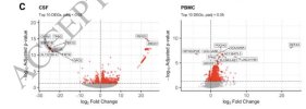
Both CSF and PBMCs demonstrate strikingly different transcriptional profiles based on NPLong COVID status. Using principal component analysis, CSF samples separated clearly based on NP-Long COVID status along PC1. In contrast, in PBMC, NP-Long COVID status did not drive separation along PC1 or PC2.


Screenshot 2025-09-14 at 3.12.23 PM copy.jpg


Screenshot 2025-09-14 at 3.10.44 PM copy.jpg
 
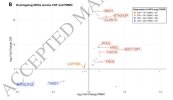
Among genes that were differentially expressed in NP-Long COVID compared to controls, there was scant overlap between DEGs in the two compartments. Of the 1,884 unique DEGs in CSF and PBMC, only 43 genes were differentially expressed in both CSF and PBMC. Furthermore, only 28 of these genes showed consistent directionality across compartments (i.e, shared direction of change in NP-Long COVID versus controls).

Using g: profiler to perform over-representation analysis, we examined the genes that were differentially expressed in both CSF and blood, but in different directions in the CSF versus the blood, when comparing NP-Long COVID to controls. These diverging genes demonstrated overrepresentation in immune-related pathways, including TNFR1-induced NF-kB signaling (genes: RACK1 and UBC) and innate immune response to SARS-CoV-1 (genes: PPIA and UBC).

Among the genes that were significantly overexpressed in the CSF of NP-Long COVID while being underexpressed in the Blood of NP- Long COVID was LAPTM5, which encodes a lysosomal transmembrane protein crucial for brain function and has been implicated in neurological diseases and immune-related processes.

Screenshot 2025-09-14 at 3.10.13 PM copy.jpg
 
Last edited:
We next identified genetic pathways that were enriched in the disease-associated gene sets by applying gene set enrichment analysis to the DEGs in the CSF and PBMC. We observed that androgen response, MTORC-related pathways, fatty acid related pathways, and protein secretion were enriched in controls (i.e, downregulated in women with NP-Long COVID) in both PBMCs and CSF.

In contrast, while reactive oxygen species and oxidative phosphorylation were both significantly enriched in PBMCs and CSF, their directionalities differ between tissues. In CSF, reactive oxygen species and oxidative phosphorylation are enriched in women with NP-Long COVID. In PBMC, however, reactive oxygen species and oxidative phosphorylation were enriched in asymptomatic women and downregulated in women with NP-Long COVID.

Several genetic pathways displayed differential expression in either CSF or PBMC of NP-Long COVID, but not both. KRAS signaling was impaired in CSF only, and MYC targets V1, notch signaling, heme metabolism, and unfolded protein response pathways were impaired in PBMCs but not CSF. Furthermore, reactive oxygen species, the P53 pathway, and oxidative phosphorylation were all enriched in the CSF of women with NP-Long COVID, but no hallmark pathways appeared to be enriched in the PBMC of women with NP-Long COVID.

there were no statistical differences in any cell type proportions in either CSF or blood
 
By analyzing the CSF compared to blood transcriptome, we identify tissue-specific immune dysregulation in the central nervous system in women with NP-Long COVID. Relative to PBMC, CSF samples demonstrate significantly more genes with large (greater than 20 fold) changes in expression level in the disease state.

pathways involved in cellular stress response, including reactive oxygen species, oxidative phosphorylation, and P53 signaling, are all enriched in the CSF of women with NP-Long COVID but not in the PBMC.

In CSF, the top upregulated gene in NP-Long COVID was FBXO2 (log2FC 22.99; p adj = 1.58 × 10⁻¹⁵ ). FBXO2 plays a critical role in waste degradation and lysophagy in the CNS.

The most downregulated gene in the CSF of NP-Long COVID was CRYM, which encodes crystallin mu (log 2 Fold Change = -26.02; p adj = 1.47 x 10⁻¹³). This crystallin, negatively regulated by T3 thyroid hormone, functions as a ketimine reductase in the brain and binds NADH or NADPH to metabolize imine bonds, and plays an important role in amino acid metabolism, particularly lysine.

Lysine is a precursor of the neurotransmitter glutamate and can exert neuroprotective effects in mammalian brains through M2 macrophage polarization . […] These results suggest that NP-Long COVID is associated with metabolic and neuroimmune alterations in the CSF compartment, including disruption of lysosomal clearance pathways and amino acid metabolism.

Interestingly, while oxidative phosphorylation appears significantly downregulated in the PBMC, we observe the opposite trend in CSF. […] These findings underscore the compartmentalized nature of NP-Long COVID and highlight the importance of evaluating CNS and systemic immune responses in parallel.

Our finding of decreased androgen response in NP-Long COVID aligns with a growing body of evidence implicating androgens in COVID-19 pathophysiology that may underlie sex differences in susceptibility

Our results also demonstrated the downregulation of fatty acid metabolism pathways in CSF samples from NP-Long COVID patients.
 
Note the over 20 fold-change up-regulation of BTN2A3P in CSF.

I wish I had a better grasp of the genetics.
Google says that BTN2A3P relates to a non-coding gene so how can it be upregulated!?

I also now note that the BTN genes are in MHC Class I. Maybe that would explain why the final DecodeME Manhattan plot does not have an MHC peak other than where BTN2A2 is?
 
I am increasingly thinking that for ME/CFS we are dealing with a failure of high-level T cell activity regulation - maybe an excessive "No" signal from regulatory or cytotoxic subpopulations - and that the female brain may be intrinsically more susceptible to knock-on effects. Maybe even there are T cell mediated events with meninges affecting CSF.
 
Completely unclear how anyone could think LC and ME/CFS are not primarily neurological disorders.

Except don't forget CSF is a lot closer to CNS tissue than blood. Apart from the blood-brain barrier, there's also proximity and dilution. So we might currently only be able to evaluate this compartment [edit: ie liquid biopsy in comparison to blood].

It's possible we might see similarly significant up/down-regulation if we sampled interstitial fluid around proximal thigh or shoulder girdle musculature or from their draining lymphatics.
 
Last edited:
If we wanted to look at this more closely or with a larger group, is lumbar puncture a procedure that could be justified in an exploratory trial?

Years ago it seemed to be regarded as risky, the sort of thing you wouldn't do unless absolutely necessary. But maybe it's more routine now? It's hard to get a sense of it.

(Of course it may not even be necessary, if as @SNT Gatchaman suggests there are other possible avenues to explore.)
 
Except don't forget CSF is a lot closer to CNS tissue than blood. Apart from the blood-brain barrier, there's also proximity and dilution. So we might currently only be able to evaluate this compartment [edit: ie liquid biopsy in comparison to blood].

It's possible we might see similarly significant up/down-regulation if we sampled interstitial fluid around proximal thigh or shoulder girdle musculature or from their draining lymphatics.

Absolutely, that wouldn't necessarily mean that it's not primarily neurological.
 
Back
Top Bottom