This post has been copied and following discussion moved from this thread: USA: NIH National Institute of Health The NIH has awarded a $2.5M R01 grant to Drs Liisa Selin and Anna Gil for their work on altered T cells in ME/CFS. This follows their 2019 Ramsay award from Solve ME with which they obtained pilot data. Press release from the Massachusetts ME/CFS Association: https://www.massmecfs.org/images/pdf/Selin_MassME_Press_Release_03162021.pdf Grant information on the NIH Project Reporter: https://reporter.nih.gov/search/-PkzeikTlEGEpI8GsfKW0A/project-details/10185392 Myalgic encephalomyelitis/chronic fatigue syndrome (ME/CFS) is a complex disorder affecting numerous organ systems and biological processes. Published data seems to suggest that ME/CFS may be preceded by infection, and the chronic manifestation of illness may represent an altered host response to infection, or an inability to resolve inflammation. Previous studies focused on perturbation in cytokines and metabolism have also shown that CD8 T responses are decreased in ME/CFS. In preliminary studies we examined the frequency, functional and phenotypic status of CD8 T cells to determine whether they were altered in chronic ME/CFS donors as compared to healthy donors. We observed an increased CD4:CD8 ratio, altered expression of exhaustion/activation markers like CTLA4 and 2B4 on CD8 T cells, and decreased production of IFN, TNF and CD107a/b upregulation following PMA stimulation, all suggesting CD8 T cell exhaustion. This was associated with a, perhaps compensatory increased frequency of activated CD4+CD8+ T cells in the ME/CFS donors as compared to healthy controls. Notably, a subset of the CD8 and the CD4+CD8+ T cell populations were spontaneously producing atypical cytokines, subdividing ME/CFS donors into two subsets: type 1 had an increased frequency of FoxP3+helios+ Treg-like cells producing IL9 (female donors); type 2 had FoxP3+helios- cells producing IL17 (male donors). When we examined the T-cell receptor (TCR) repertoire of the CD4+CD8+ T cell population we found evidence of antigen driven clonal expansions to an unknown antigen at this time, whether it will be a viral or auto-antigen. We hypothesize that the common theme in ME/CFS is an aberrant response to an immunological trigger like infection, which results in a permanently dysregulated immune system as a result of CD8 T cell exhaustion. These studies will identify potential biomarkers and mechanisms driving the immunopathogenesis of ME/CFS leading to future therapies. We will explore this hypothesis in the following Aims. Aim 1 we will examine altered CD8 and CD4+CD8+ T-cell responses in ME/CFS: 1) we will determine if the level of CD8 T cell exhaustion varies with ME/CFS type 1 (female) and Type 2 (male) response and with the severity of ME/CFS symptoms using a larger ME/CFS cohort; 2) we will examine EBV antigen-specific responses in ME/CFS donors to determine if a common persistent virus response is altered by the immunosuppressive state of CD8 T cell exhaustion and further contributing to the disease state of ME/CFS; 3) microarray analyses will be done on sorted activated T cell subsets to assist in understanding the alterations in the functionality of the exhausted and activated CD8 and CD4+CD8+ T cell subsets in ME/CFS donors. In Aim 2 we will examine TCR repertoire of CD8 and CD4+CD8+ T-cell subsets for evidence of antigen driven clonal expansion. Defining the characteristics of the activated clonally expanded CD8 and CD4+CD8+ T cells would be a major step in the field potentially leading to the identification a specific infectious or auto-antigen response that could be the main driver of CD8 T cell exhaustion and the immunological basis of ME/CFS. ETA: The MassME press release links to a presentation from September 2020 and a conference poster with Seliin and Gil's preliminary findings.
Have these been discussed yet? Looks like the sort of thing that should have been but can't find any threads? Just pilot data, tiny numbers, no detail on cohort selection given etc. etc. but they do seem to be rather intriguing early findings (at least if like me you don't fully understand them). The funders must have thought there was something there since they've given this big grant based on them. What do those of you who actually understand T-cells properly think? The video discusses the graphs on the poster. Fluctuating sound quality but worth the effort.
This thread Severity of Acute Infectious Mononucleosis Correlates with Cross-Reactive Influenza CD8 T-Cell Receptor Repertoires Nuray Aslan et al 2017 discuss Selin and Gil's work. It's also mentioned here: The possibility of autoimmunity or auto-reactivity in ME/CFS (edited to take into account the new thread)
Didn't fluge and mella target t-cells with mixed results? I never followed that line of research closely.
Moved post The conference poster and a video presentation with some interesting pilot data which haven't been discussed yet (I don't think): https://www.massmecfs.org/images/pdf/MECFS_FOCIS_poster_2020.pdf video presentation related to above poster: Abstract and summary taken from the conference poster: "Abstract Myalgic Encephalomyelitis/ChronicFatigueSyndrome (ME/CFS) is a complex disorder affecting numerous organ systems and biological processes. Published data seems to suggest that ME/CFS may be preceded by infection, and the chronic manifestation of illness may represent an altered host response to infection, or an inability to resolve inflammation. Previous studies focused on perturbation in cytokines and metabolism have shown that CD8+ T responses are decreased in ME/CFS. Here, we hypothesize that in ME/CFS an aberrant response to an immunological trigger like infection may result in a permanently dysregulated immune system, leading to a state of immunosuppression. We examined the frequency, functional and phenotypic status of CD8+ and CD4+CD8+ T-cells to determine whether their frequency and cytokine production was altered in chronic ME/CFS patients (ME/CFS) as compared to healthy donors (HDs). We examined the T-cell receptor (TCR) repertoire of the CD4+CD8+ population looking for evidence consistent with an antigendriven response whether it will be a viral or auto-antigen. We observed altered expression of exhaustion markers like CTLA4 and 2B4, decrease in CD8 T-cell number, and function, particularly CD107ab and IFNg production. This was associated with a compensatory increased frequency of activated CD4+CD8+ Tcells in ME/CFS patients as compared to healthy controls. Both the CD8 and CD4+CD8+ Tcell populations were spontaneously producing aberrant cytokines, subdividing into two types of ME/CFS: (1) FoxP3+ cells producing IL9 (female donors), (2) IL17-producing cells (male donors). TCR analyses suggested an antigen-driven response. These results are consistent with immunosuppression mediated via exhaustion of CD8 T-cells as observed either in chronic viral infections or tumor environments. The observed exhaustion was associated with a compensatory increase in activated CD4+CD8+ that make unusual cytokines known to interact with the nervous system. These findings identify potential biomarkers and mechanisms driving the immunopathogenesis of ME/CFS leading to future therapies (Funding: Ramsay Award, SolveME/CFS Initiative). Summary 1. Increased frequency of CD4+CD8+ T cells and low CD8 T cell frequency in ME/CFS donors as compared to healthy controls (potential biomarker). The CD4+CD8+ cells are producing cytokines without stimulation and express high levels of CTLA4. (What is driving their activation?) 2. Evidenced of exhausted CD8 T cells in ME/CFS donors: Decreased production of cytokines (IFNg, MIP1b and TNFa) after ex vivo stimulation in ME/CFS donors (potential biomarker). Decreased expression of 2B4 on CD8 T cells Increased expression of CTLA4 Is there a persistent antigen (virus or autoantigen?) 3. TCRαβ repertoire analysis of CD4+CD8+ T cells demonstrated highly polyclonal response with features indicating an antigen-driven response (persistent virus or autoantigen?) 4. Dysregulated unusual cytokine responses in CD4+CD8+ and subset of CD8 T cells of ME/CFS donors without any stimulation: IL-9in females or IL-17 in males)."
Thanks. I've posted the conference poster and video links in the Severity of Acute Infectious Mononucleosis Correlates with Cross-Reactive Influenza CD8 T-Cell Receptor Repertoires Nuray Aslan et al 2017 thread for further discussion there. I hope. Really would like to know what others think.
Merged thread https://twitter.com/user/status/1371995912877264897 Altered T-cell response in ME/cfs https://www.massmecfs.org/images/pdf/Selin_MassME_Press_Release_03162021.pdf This NIH RO1 grant, titled “Altered T cell Responses in ME/CFS” allows the researchers to examine the role of aberrant T cell responses in the immunopathogenesis of ME/CFS patients. Selin and Gil's recent research findings could point to potential biomarkers, treatments and ways of tracking response to therapy for the disease, things that have been sorely missing. To date, there is no FDA approved treatment for ME/CFS, a devastating disease with neurological and immunological characteristics, afflicting up to 2.5 million Americans.
Hopefully, this NIH grant will encourage other researchers to submit ME/CFS related grant applications. I realize that the NIH does not have a great history approving such applications - and they bemoan not getting enough high quality submissions - but that is at least, in part, a chicken and egg situation. If they want more people to submit high quality applications, there has to be a perception that it's worth the time and effort to do so, otherwise it looks like the NIH simply does not approve ME/CFS related applications. In economic terms I believe this called "priming the pump," a metaphor for spending an initial amount of money just to get things moving.
They made a presentation recently on their work, here is the video (new to me, not sure if it’s posted somewhere else on the forum). Quite fascinating but there were just a few patients tested, therefore a larger cohort will be telling in determining whether there is a there there.
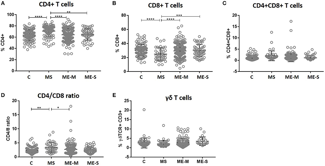
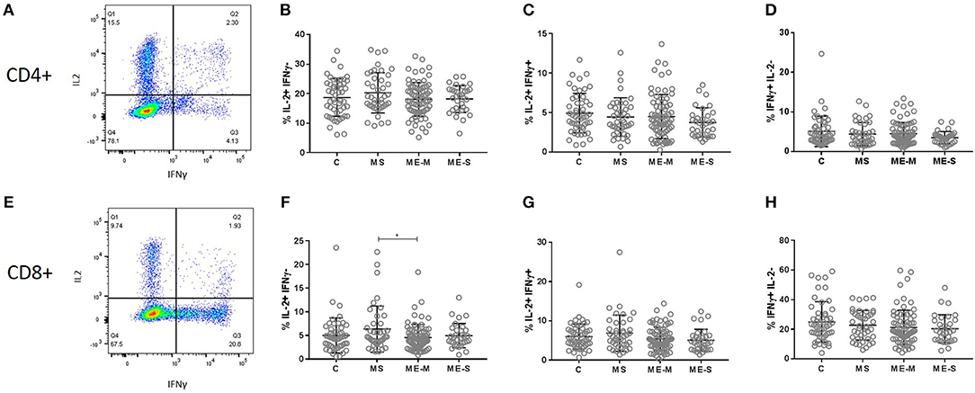
I'm afraid that some of Gil and Seliin's findings may be due to their small sample size (10 ME/CFS patients, 16 healthy controls). The UK ME/CFS Biobank's 2019 immunology study (251 ME/CFS of which 54 were severe, 107 healthy controls, 46 MS controls) did not find a difference between ME/CFS and HC donors in CD4+, CD8+ or CD4+CD8+ T cell counts, nor for the CD4/CD8 ratio: Figure 3. T cell subset quantification in PBMC from people with ME/CFS, people with MS and healthy controls. Within the T cell gate (CD3+), the CD4+ and CD8 staining was characterised to calculate the proportion of (A) CD4+ T cells, (B) CD8+ T cells, (C) double positive CD4+CD8+ T cells, and (D) the ratio of CD4+:CD8+ T cells. (E) The proportion of T cells which expressed the γδ TCR within the CD3+ T cell population were determined. Data are from healthy controls (C: n = 107), multiple sclerosis (MS: n = 46), mild/moderate ME/CFS (ME-M: n = 197), and severely affected ME/CFS (ME-S: n = 54) for (A–D), and from C (n = 56), MS (n = 46), ME-M (n = 120), and ME-S (n = 21) for (E). Clinical groups were compared by Kruskal-Wallis test for non-parametric data: *P < 0.05, **P < 0.01, ***P < 0.001, ****P < 0.0001. Within the lymphocyte gate, the proportions of cells that were either CD4+ T cells or CD8+ T cells (and, as a consequence, the CD4/CD8 ratio) differed significantly between groups in the unadjusted analysis (ANOVA P < 0.0001 in each case) and these differences persisted after adjusting for confounders (P < 0.0001 in each case) (Figure 3). However, these differences were entirely due to significantly higher proportions of CD4+ T cells, lower proportions of CD8+ T cells and thus a higher CD4/CD8 ratio in the MS patients; there were no significant differences in lymphocyte distribution between ME/CFS cases and healthy controls. Similarly, there were no significant differences between the groups in the absolute numbers of different leucocyte and lymphocyte populations (Supplementary Figure S2). There were no differences either in the production of IFN-gamma by CD8+ T cells (comparing this figure to Figure 6 of Gil and Seliin's poster): Figure 8. Production of cytokines by T cells in response to stimulation in vitro. PBMC from mild/moderately affected ME/CFS (ME-M: n = 76), severely affected ME/CFS (ME-S: n = 32) or multiple sclerosis (MS: n = 41) patients or healthy control (C: n = 50) individuals were cultured in vitro with PMA and ionomycin for 4 h. The production of IFNγ and IL-2 cytokines was assessed in CD4+ (A–D) and CD8+ (E–H) T cells, with the proportions of cells which produced only IL2 (B,F), both IL2 and IFNγ (C,G) or only IFNγ (D,H) calculated for each study participant. Within the dot plots, the lines show the means and the error bars show ± SD. [...] The proportion of cells producing IL-2, IFN-γ or a combination of IL-2 and IFN-γ was assessed in the CD3+ CD4+ and the CD3+ CD8+ cell populations but no significant differences were observed among the groups in either the adjusted or unadjusted analysis (Figure 8).
It might be that while the subset ratios and total counts don't vary between ME/CFS patients and controls, the functioning of those T Cells does vary. T Cell exhaustion is something of a trendy research area, but that is not such a bad thing. Here is some research about mitochondria of T Cells in HIV: Frontiers | Mitochondrial functions are compromised in CD4 T cells from ART-controlled PLHIV | Immunology (frontiersin.org). The good thing about the HIV analogy, really, I think is that the impacts of HIV on the immune system are highly complex, but that complex knowledge over time has resulted in treatment approaches. Seems to me, also, that the onset event of ME/CFS often follows a long period of flu-like symptoms. But what happens at the point of that 'onset event' is not known.
Could some of the different CD4+CD8+ findings be due to the method used to count them? Gil and Seliin didn't find any increased proportion in 'unsorted' blood (whatever that is) and only found increased proportion after doing magnet sorting (whatever that is). The Biobank study doesn't mention magnet sorting so presumably didn't use it. Magnetic sorting still wouldn't explain the different findings in the CD4/CD8 ratios because Gil and Seliin calculated those based on the unsorted blood results. But there's a slide at about 22 minutes into the video where they looked at 1 patient and 1 control over a long period, more than a decade, and found the unsorted results in the patient are very up and down so the time of sampling could have a large effect on results, especially in tiny cohorts. The control was much more stable and both were quite stable for the magnet sorted counts. I don't know what any of this means. But yes, the low numbers are a worry. Though it looks like this is an ongoing study so a more definite picture may yet emerge.
This press release seems to be linked to the study discussed in this thread (it has the same NIH grant number). https://www.unomaha.edu/news/2022/02/ghersi-immune-system-research.php
The #MEAction Network New T-Cell Research At The University Of Massachusetts! Read more here: https://www.meaction.net/2022/05/18/t-cell-umass/ #MEAction’s Director of Scientific and Medical Outreach, Jaime Seltzer, sat down with Dr. Liisa Selin, MD, PhD, and Dr. Anna Gil, PhD, recipients of an R01 grant to research immune system dysregulation in people with ME/CFS. Selin, a professor of pathology and a person with ME herself, and Gil, a viral immunologist, comprise the two-person Selin lab at the University of Massachusetts T.H. Chan Medical School. Selin and Gil’s primary area of study involves T cells, white blood cells involved in the immune response to infection. Check out the article for more info! Recently, the Selin Lab helped organize a meeting with Congressmen Jim McGovern (D-MA) and Jamie Raskin (D-MD) between researchers and advocates for ME and Long COVID at the U Mass Chan Medical School. Both Selin and Gil spoke about their research.
Sorting means selecting/filtering a subset of cells based on the expression of some antigen. In this case it's CD3 which is exclusively found on T cells with very few exceptions - on the other hand CD4 and CD8 marker are also found on monocytes, macrophages, natural killer cells, dendritic cells and probably others..
They have a new website: https://www.umassmed.edu/selinlab/ 10-item Twitter thread Can be read here: https://threadreaderapp.com/thread/1836068503490638018.html As the website says, "Dr. Selin has ME/CFS".