Transdiagnostic reduction in cortical choline-containing compounds in anxiety disorders: a 1H-magnetic resonance spectroscopy meta-analysis
Maddock, Richard J.; Smucny, Jason
[Line breaks added]
Background
Anxiety disorders (AnxDs) are highly prevalent and often untreated or unresponsive to treatment. Although proton magnetic resonance spectroscopy (1H-MRS) studies of AnxDs have been conducted for over 25 years, a consensus regarding neurometabolic abnormalities in these conditions is lacking.
Methods
A systematic review and meta-analysis of 1H-MRS studies of AnxDs (social anxiety disorder, generalized anxiety disorder, and panic disorder) identified 25 published datasets meeting inclusion criteria. These compared neurometabolites between 370 patients and 342 controls, including n-acetlyaspartate (NAA), total creatine, total choline (tCho), myo-inositol, glutamate, glutamate+glutamine, GABA, and lactate.
Results
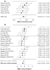
Across AnxDs, tCho was significantly reduced in prefrontal cortex and across all cortical regions. Effect sizes for cortical tCho were significantly more negative in studies with better measurement quality, with Hedges’ g = −0.64 and an 8% mean reduction.
NAA was unchanged in prefrontal cortex but reduced across all cortical regions (after exclusions).
These abnormalities did not differ between the three disorders. No other neurometabolites differed significantly.
Discussion
Reduced choline-containing compounds in cortical regions is a consistent, transdiagnostic abnormality in AnxDs.
Notably, arousal-related neuromodulators, including norepinephrine, alter membrane phospholipid homeostasis and methylation reactions, which influence brain tCho levels. This suggests that chronically elevated arousal in AnxDs may increase neurometabolic demand for choline compounds without a proportionate increase in brain uptake, leading to reduced tCho levels.
Reduced cortical NAA suggests compromised neuronal function in AnxDs.
Future studies may clarify the clinical significance of reduced cortical tCho and the possibility that appropriate choline supplementation could have therapeutic benefit in anxiety disorders.
Web | PDF | Molecular Psychiatry | Open Access

