Preprint Biological Insights from Genome-Wide Association Studies and Whole Genome Sequencing of [ME/CFS], 2026, Maccallini et al

forestglip

Administrator
Staff member
Biological Insights from Genome-Wide Association Studies and Whole Genome Sequencing of Myalgic Encephalomyelitis/Chronic Fatigue Syndrome

Maccallini, Paolo

Abstract
Myalgic encephalomyelitis/chronic fatigue syndrome (ME/CFS) is a debilitating disorder of poorly understood etiology.

We performed a meta-analysis of two European-ancestry ME/CFS genome-wide association studies (GWAS) with no overlap in subjects, DecodeME and Million Veteran Program, comprising a total of 19,470 cases and 699,111 controls. Post-GWAS analyses investigated the association between ME/CFS and specific tissues, cell types, cellular components, and canonical pathways. Findings were independently evaluated for replication against a module of ME/CFS risk genes previously prioritized by machine learning applied to rare coding variants from whole-genome sequencing (WGS) of ME/CFS cases and controls of European ancestry.

Tissue enrichment analysis implicated multiple brain regions and the pituitary, with no peripheral tissue reaching significance, and three survived Bonferroni-corrected replication. Gene-set analysis identified multiple neuronal and synaptic gene sets, several of which were independently replicated, with glutamatergic synapses as the most specific replicated signal. Cell-type analysis identified independent replicated signals in distinct neuronal populations of subcortical and cerebellar regions.

These results suggest a role for synaptic function in specific brain regions in the pathogenesis of ME/CFS, with convergent support from both common and rare variant data. Larger studies are needed to confirm these findings and to identify specific targets for therapeutic intervention.

Web | DOI | Zenodo | Preprint
 
This is impressive work and well worth an in depth-read!

It follows the same line of reasoning we've been talking about on the forum fore more than a year, based on both the DecodeME data on common mutations and Mark Synder study on rare mutations hinting at neural synapses in the pathology of ME/CFS. But Paolo has done it in a more formal and persuasive way.

A first important addition is that he managed to add the 3891 ME/CFS cases from the Million Veteran Program (MVP) to the 15,579 from DecodeME in a meta-analysis. Paolo said he wanted to add cases from other genetic databases as well such as the UK biobank or FinnGenn, but that gave the issue that some of the controls overlapped. So this (DecodeME + MVP) is the biggest meta-analysis he could do.

Those from MVP were based on ICD-codes and with a prevalences of around 1.5% they might contain a lot of false positives (patients label ME/CFS who might not have it). But the bump in statistical power was just enough to get a lot of interesting results.

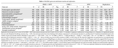
Paolo used the MAGMA tool that uses database on gene expression to see if the DNA results from ME/CFS patients are enriched in a particular group of genes or tissue. The DecodeME preprint did this as well and we (mostly forestglip) played around with this on the forum as well, showing it mostly pointed to the brain. But with the added MVP samples, Paolo seem to have gotten much stronger result implicating synapses in multiple ways.

1778872216793.png
The second interesting thing he added is that he used these gene sets from MAGMA and tested them in the Snyder study that used rare mutations. Several of these gene sets were replicated. Here are the important paragraphs that describe this approach:
Gene-set analysis using MAGMA tested 17,009 of the 17,023 gene sets in the input file(MSigDB_20231Hs_MAGMA.txt). Twelve gene sets were significantly enriched for association signalin the DME-1 + MVP meta-analysis after Benjamini-Hochberg correction (PBH < 0.05, k = 17,009;Table 4). All 12 span neuronal and synaptic categories from Gene Ontology cellular components,Gene Ontology molecular functions, and Reactome. Three of the 12 also survived the more stringent Bonferroni correction (P < 0.05/17,009 = 2.94 × 10⁻⁶): postsynaptic specialization, neuron-to-neuronsynapse, and somatodendritic compartment, all Gene Ontology cellular component terms implicating the synapse in general and post-synapses in particular. Neither DME-1 nor MVP alone reached significant enrichment after BH correction (S1 File, sheet S3).
Replication was evaluated using the HEAL2 module of ME/CFS risk genes [20]. Only 6,063 of the17,023 input gene sets had non-empty overlap with the HEAL2 background (17,759 genes) and were testable. Seven of the 12 BH-significant gene sets were replicated at Bonferroni-corrected significance(P < 0.05/12 = 4.17 × 10⁻³; shaded rows in Table 4), of which six also survived Bonferroni correctionin the HEAL2 module (P < 0.05/6,063 = 8.25 × 10⁻⁶; bold replication p-values in Table 4). All three gene sets surviving Bonferroni correction in discovery were replicated at Bonferroni-correctedsignificance (P < 0.05/3 = 0.017; P†in Table 4).
I think this is quite big, because it's not just about DecodeME (one study anymore) or even one approach (GWAS of common alleles).

The tissue results were similar to those from DecodeME alone I believe (various brain regions), but using various gene expression databases, Paolo found significant hits for particular brain cell types, most notably:

- the eccentric medium spiny neuron (eMSN) in the claustrum
- glutamatergic neuron in cerebellar white matter
 
Last edited:
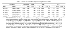
These were the 5 hits in the meta-analysis. The one on chromosome 17 for CA10 is not there but we do have a new one on chromosome 19.
1778874942972.png

Paolo provided the GRCh38 location in the supplementary material:

topLeadSNPGRCh38GRCh37
rs25037736:98089269:A:G6:98537145:A:G
rs1257972212:118125397:T:C12:118563202:T:C
rs716532715:54866724:A:G15:55158922:A:G
rs198293519:31366884:C:T19:31857790:C:T
rs481089420:48916462:A:G20:47532999:A:G

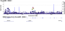
When I looked up the location on chr 19 on LocusZoom I got the following picture:

1778875129662.png

TSHZ3 seems to be involved in neural development.
 
I wonder if people with ME/CFS want to be thought of as medium spiny and in the claustrum?
There have been so many weird and denigrating names for our disease: chronic fatigue syndrome, post-exertional malaise, sickness behavior, photophobia.

If anything it must mean we're on the right track with "eccentric medium spiny neuron (eMSN) in the claustrum"!
 
The cerebellum has been mentioned in several places .The cerebellum is a major sensory processing center. It maintains upright posture ?OI
It fine tunes motor coordination and we have problems with ?handwriting. It is also involved with blood pressure regulation.

 
I have been talking with Paolo for quite some time. One of our talks was -given these results and specificly glutamatergic synapses- as to what could be the role of glutamate and what kind of interventions could be used.He mentioned a specific medication. I do not know whether Paolo would like this discussion to be shared, I will ask him to comment or whether it is ok to be shared.

I am not an expert but would the use of H-MAGMA be a next analysis worth investigating?

I am also *deeply* concerned because I get the impression that ME/CFS is beginning to appear as a "brain disorder". Having clearer signals pointing to the brain does not necessarily mean that ME/CFS is a brain-related disease.
 
Last edited:
I am also *deeply* concerned because I get the impression that ME/CFS is beginning to appear as a "brain disorder". Having clearer signals pointing to the brain does not necessarily mean that ME/CFS is a brain-related disease.

ME/CFS has pretty much always appeared as a brain-related disorder. It is a matter of clinical fact - with sensory sensitivity, sleep disturbance, cognitive change etc. Simon, Adrian, Mark, Andrew and I said so in 2016 in Fatigue and Biomedicine before any genes had been found.

The brain may be the target of systemic shifts arising from other organ systems. But the age of onset, and unpredictable waxing and waning make an adaptive immune responsive (maybe an a rather wide sense) about the only thing I can think of with the right dynamics.

I don't think we should be concerned that the data seem to fit.
And Paolo's angle looks very plausible.
 
ME/CFS has pretty much always appeared as a brain-related disorder. It is a matter of clinical fact - with sensory sensitivity, sleep disturbance, cognitive change etc. Simon, Adrian, Mark, Andrew and I said so in 2016 in Fatigue and Biomedicine before any genes had been found.

The brain may be the target of systemic shifts arising from other organ systems. But the age of onset, and unpredictable waxing and waning make an adaptive immune responsive (maybe an a rather wide sense) about the only thing I can think of with the right dynamics.

I don't think we should be concerned that the data seem to fit.
And Paolo's angle looks very plausible.
Are you suggesting that the cause of ME/CFS is brain-related?
 
Are you suggesting that the cause of ME/CFS is brain-related?

I think the 'cause' must lie in some inherent instability in the complex regulatory systems of the human body. In a sense the cause of a disease is always the totality of the human condition and its environment. And since evolution re-writes genes at random the 'program' for this totality is what Gary Marcus called a 'kluge' - a mess of things that happen to work but make use of all sorts of random overlapping tricks.

But, to narrow that down, what we implied in 2016 was that the instability was most likely to be in the brain set up, or the immune system or both. That does not exclude mediation through all sorts of other pathways, and involvement of other tissues, as occurs in the wide spectrum of autoimmune diseases. But so far, I have not seen consistent evidence of any pathology in other tissues.
 
I think the 'cause' must lie in some inherent instability in the complex regulatory systems of the human body. In a sense the cause of a disease is always the totality of the human condition and its environment. And since evolution re-writes genes at random the 'program' for this totality is what Gary Marcus called a 'kluge' - a mess of things that happen to work but make use of all sorts of random overlapping tricks.

But, to narrow that down, what we implied in 2016 was that the instability was most likely to be in the brain set up, or the immune system or both. That does not exclude mediation through all sorts of other pathways, and involvement of other tissues, as occurs in the wide spectrum of autoimmune diseases. But so far, I have not seen consistent evidence of any pathology in other tissues.
I see, so you are effectively leaving out of the picture the numerous metabolic disturbances which we are seeing, unless you believe that these disturbances are either erroneous or completely irrelevant. Just making sure because making assumptions could have a negative outcome here.
 
I see, so you are effectively leaving out of the picture the numerous metabolic disturbances which we are seeing, unless you believe that these disturbances are either erroneous or completely irrelevant.

I am not seeing any metabolic disturbances outside the normal range. There may be some statistical shifts in some studies but what came up in the Beentjes study looked likely to be epiphenomenal or confounding to me. Do you see any consistent metabolic shifts?

I agree with Snow Leopard that the second day CPET looks as if it may be an effect mediated by muscle afferents nerves.
 
And since evolution re-writes genes at random the 'program' for this totality is what Gary Marcus called a 'kluge' - a mess of things that happen to work but make use of all sorts of random overlapping tricks.

Eg where synapse genes (esp. glutamatergic) may be affecting B and T cell receptors —

The Scaffolding Protein Synapse-Associated Protein 97 Is Required for Enhanced Signaling Through Isotype-Switched IgG Memory B Cell Receptors (2012, Science Signaling)
After their first encounter with a foreign antigen, naive B cells that have immunoglobulin M (IgM) B cell receptors (BCRs) trigger the primary antibody response and the generation of memory B cells with IgG BCRs. When these memory B cells reencounter the same antigen, the cell surface IgG BCRs stimulate their rapid differentiation into plasma cells that release large amounts of IgG antibodies.

We showed that the conserved cytoplasmic tail of the IgG BCR, which contains a putative PDZ (postsynaptic density 95/disc large/zona occludens 1)–binding motif, associated with synapse-associated protein 97 (SAP97), a PDZ domain–containing scaffolding molecule that is involved in controlling receptor density and signal strength at neuronal synapses. SAP97 accumulated and bound to IgG BCRs in the immunological synapses that formed in response to B cell engagement with antigen. Knocking down SAP97 in IgG+ B cells or mutating the putative PDZ-binding motif in the BCR tail impaired formation of the immunological synapse, initiation of IgG BCR signaling, and downstream activation of the mitogen-activated protein kinase p38.

Thus, heightened B cell memory responses are encoded, in part, by a mechanism that involves SAP97 serving as a scaffolding protein in the IgG BCR immunological synapse.

The role of PSD-95 in the rearrangement of Kv1.3 channels to the immunological synapse (2013, Pflügers Archiv - European Journal of Physiology)
Establishment of the immunological synapse (IS) between T lymphocytes and antigen-presenting cells is a key step in the adaptive immune response. Several proteins accumulate in the IS, such as the Kv1.3 potassium channel; however, the mechanism of this translocation is unknown.

PSD-95 and SAP97 are adaptor proteins that regulate the polarized cell surface expression and localization of Kv1 channels in neurons. We investigated whether these proteins affect the redistribution of Kv1.3 into the IS in non-excitable human T cells. We show here that PSD-95 and SAP97 are expressed in Jurkat and interact with the C terminus of Kv1.3.

Disruption of the interaction between PSD-95 or SAP97 and Kv1.3 in Jurkat was realized by the expression of a C-terminal truncated Kv1.3, which lacks the binding domain for these proteins, or by the knockdown of the expression of PSD-95 or SAP97 using specific shRNA. Expression of the truncated Kv1.3 or knockdown of PSD-95, but not the knockdown of SAP97, inhibited the recruitment of Kv1.3 into the IS; the fraction of cells showing polarized Kv1.3 expression upon engagement in an IS was significantly lower than in control cells expressing the full-length Kv1.3, and the rearrangement of Kv1.3 did not show time dependence. In contrast, Jurkat cells expressing the full-length channel showed marked time dependence in the recruitment into the IS peaking at 1 min after the conjugation of the cells.

These results demonstrate that PSD-95 participates in the targeting of Kv1.3 into the IS, implying its important role in human T-cell activation.

The neurotransmitter glutamate and human T cells: glutamate receptors and glutamate-induced direct and potent effects on normal human T cells, cancerous human leukemia and lymphoma T cells, and autoimmune human T cells (2014, Journal of Neural Transmission)
Glutamate is the most important excitatory neurotransmitter of the nervous system, critically needed for the brain’s development and function. Glutamate has also a signaling role in peripheral organs.

Herein, we discuss glutamate receptors (GluRs) and glutamate-induced direct effects on human T cells. T cells are the most important cells of the adaptive immune system, crucially needed for eradication of all infectious organisms and cancer. Normal, cancer and autoimmune human T cells express functional ionotropic and metabotropic GluRs. Different GluR subtypes are expressed in different T cell subtypes, and in resting vs. activated T cells.

Glutamate by itself, at low physiological 10−8M to 10−5M concentrations and via its several types of GluRs, activates many key T cell functions in normal human T cells, among them adhesion, migration, proliferation, intracellular Ca2+ fluxes, outward K+ currents and more. Glutamate also protects activated T cells from antigen-induced apoptotic cell death. By doing all that, glutamate can improve substantially the function and survival of resting and activated human T cells. Yet, glutamate’s direct effects on T cells depend dramatically on its concentration and might be inhibitory at excess pathological 10−3M glutamate concentrations.

The effects of glutamate on T cells also depend on the specific GluRs types expressed on the target T cells, the T cell’s type and subtype, the T cell’s resting or activated state, and the presence or absence of other simultaneous stimuli besides glutamate. Glutamate also seems to play an active role in T cell diseases. For example, glutamate at several concentrations induces or enhances significantly very important functions of human T-leukemia and T-lymphoma cells, among them adhesion to the extracellular matrix, migration, in vivo engraftment into solid organs, and the production and secretion of the cancer-associated matrix metalloproteinase MMP-9 and its inducer CD147. Glutamate induces all these effects via activation of GluRs highly expressed in human T-leukemia and T-lymphoma cells. Glutamate also affects T cell-mediated autoimmune diseases.

With regards to multiple sclerosis (MS), GluR3 is highly expressed in T cells of MS patients, and upregulated significantly during relapse and when there is neurological evidence of disease activity. Moreover, glutamate or AMPA (10−8M to 10−5M) enhances the proliferation of autoreactive T cells of MS patients in response to myelin proteins. Thus, glutamate may play an active role in MS. Glutamate and its receptors also seem to be involved in autoimmune rheumatoid arthritis and systemic lupus erythematosus. Finally, T cells can produce and release glutamate that in turn affects other cells, and during the contact between T cells and dendritic cells, the latter cells release glutamate that has potent effects on the T cells.

Together, these evidences show that glutamate has very potent effects on normal, and also on cancer and autoimmune pathological T cells. Moreover, these evidences suggest that glutamate and glutamate-receptor agonists might be used for inducing and boosting beneficial T cell functions, for example, T cell activity against cancer and infectious organisms, and that glutamate-receptor antagonists might be used for preventing glutamate-induced activating effects on detrimental autoimmune and cancerous T cells.

Glutamate receptor–T cell receptor signaling potentiates full CD8+ T cell activation and effector function in tumor immunity (2025, iScience)
Glutamate is best known as an excitatory neurotransmitter. However, its roles in T cell immunity remain underrecognized. We investigated the interplay between glutamate receptors (GluRs) and T cell receptors (TCRs) during CD8+ T cell activation. Our findings reveal that GluR expression in CD8+ T lymphocytes strongly correlates with the activation of TCR-CD28 signaling, enhancing their antitumor effector responses.

Conversely, pharmacologic antagonism of GluRs in activated CD8+ T cells disrupts the colocalization of GluR with TCRVβ8.1, reduces the phosphorylation of TCR-signaling intermediates, alters calcium flux, and impairs the metabolic switch to glycolysis essential for T cell activation. Moreover, these disruptions blunt clonal proliferation and compromise the tumor-cytolytic capacity of CD8+ T cells.

Thus, the glutamatergic system—via the GluR−TCR signaling complex—plays a critical amplifier role in activating CD8+ T cells and eliciting their full antitumor activity. This mechanistic insight reveals a previously underappreciated axis in T cell biology and opens avenues for immunotherapy regimens targeting GluR−TCR interactions to augment T cell–mediated responses in cancer and potentially other immunopathologies.

HIGHLIGHTS
• Expression of GluR, CD69 and CD25 is critical for the activation of CD8+ T lymphocytes

• Inhibiting GluR signaling blunts CD8+ T cell proliferation, glycolysis and effector function

• Activation of the GluR−TCR−CD28 axis elicits full CD8+ T cell antitumor effector responses

• Engagement of mGluR1 shapes T cell immunity in the solid tumor microenvironment

Or vascular function —

The synaptic proteins neurexins and neuroligins are widely expressed in the vascular system and contribute to its functions (2009, Proceedings of the National Academy of Sciences)
Unlike other neuronal counterparts, primary synaptic proteins are not known to be involved in vascular physiology. Here, we demonstrate that neurexins and neuroligins, which constitute large and complex families of fundamental players in synaptic activity, are produced and processed by endothelial and vascular smooth muscle cells throughout the vasculature. Moreover, they are dynamically regulated during vessel remodeling and form endogenous complexes in large vessels as well as in the brain.

We used the chicken chorioallantoic membrane as a system to pursue functional studies and demonstrate that a monoclonal recombinant antibody against β-neurexin inhibits angiogenesis, whereas exogenous neuroligin has a role in promoting angiogenesis. Finally, as an insight into the mechanism of action of β-neurexin, we show that the anti-β-neurexin antibody influences vessel tone in isolated chicken arteries.

Our finding strongly supports the idea that even the most complex and plastic events taking place in the nervous system (i.e., synaptic activity) share molecular cues with the vascular system.

Neurexins and neuroligins: synapses look out of the nervous system (2011, Cellular and Molecular Life Sciences)
The scientific interest in the family of the so-called nervous vascular parallels has been growing steadily for the past 15 years, either by addition of new members to the group or, lately, by deepening the analysis of established concepts and mediators. Proteins governing both neurons and vascular cells are known to be involved in events such as cell fate determination and migration/guidance but not in the last and apparently most complex step of nervous system development, the formation and maturation of synapses.

Hence, the recent addition to this family of the specific synaptic proteins, Neurexin and Neuroligin, is a double innovation. The two proteins, which were thought to be “simple” adhesive links between the pre- and post-synaptic sides of chemical synapses, are in fact extremely complex and modulate the most subtle synaptic activities. We will discuss the relevant data and the intriguing challenge of transferring synaptic activities to vascular functions.

NMDA-Type Glutamate Receptor Activation Promotes Vascular Remodeling and Pulmonary Arterial Hypertension (2018, Circulation)
BACKGROUND
Excessive proliferation and apoptosis resistance in pulmonary vascular cells underlie vascular remodeling in pulmonary arterial hypertension (PAH). Specific treatments for PAH exist, mostly targeting endothelial dysfunction, but high pulmonary arterial pressure still causes heart failure and death. Pulmonary vascular remodeling may be driven by metabolic reprogramming of vascular cells to increase glutaminolysis and glutamate production. The N-methyl-d-aspartate receptor (NMDAR), a major neuronal glutamate receptor, is also expressed on vascular cells, but its role in PAH is unknown.

METHODS
We assessed the status of the glutamate-NMDAR axis in the pulmonary arteries of patients with PAH and controls through mass spectrometry imaging, Western blotting, and immunohistochemistry. We measured the glutamate release from cultured pulmonary vascular cells using enzymatic assays and analyzed NMDAR regulation/phosphorylation through Western blot experiments. The effect of NMDAR blockade on human pulmonary arterial smooth muscle cell proliferation was determined using a BrdU incorporation assay. We assessed the role of NMDARs in vascular remodeling associated to pulmonary hypertension, in both smooth muscle-specific NMDAR knockout mice exposed to chronic hypoxia and the monocrotaline rat model of pulmonary hypertension using NMDAR blockers.

RESULTS
We report glutamate accumulation, upregulation of the NMDAR, and NMDAR engagement reflected by increases in GluN1-subunit phosphorylation in the pulmonary arteries of human patients with PAH. Kv channel inhibition and type A-selective endothelin receptor activation amplified calcium-dependent glutamate release from human pulmonary arterial smooth muscle cell, and type A-selective endothelin receptor and platelet-derived growth factor receptor activation led to NMDAR engagement, highlighting crosstalk between the glutamate-NMDAR axis and major PAH-associated pathways. The platelet-derived growth factor-BB-induced proliferation of human pulmonary arterial smooth muscle cells involved NMDAR activation and phosphorylated GluN1 subunit localization to cell-cell contacts, consistent with glutamatergic communication between proliferating human pulmonary arterial smooth muscle cells via NMDARs. Smooth-muscle NMDAR deficiency in mice attenuated the vascular remodeling triggered by chronic hypoxia, highlighting the role of vascular NMDARs in pulmonary hypertension. Pharmacological NMDAR blockade in the monocrotaline rat model of pulmonary hypertension had beneficial effects on cardiac and vascular remodeling, decreasing endothelial dysfunction, cell proliferation, and apoptosis resistance while disrupting the glutamate-NMDAR pathway in pulmonary arteries.

CONCLUSIONS
These results reveal a dysregulation of the glutamate-NMDAR axis in the pulmonary arteries of patients with PAH and identify vascular NMDARs as targets for antiremodeling treatments in PAH.
 
Topic Glutamate: I’m currently testing a medication that inhibits the release of neurotransmitters such as glutamate. It’s still far too early to say anything about its effectiveness, but two days ago I was able to watch half a football match on my phone. Before starting the medication, that had no longer been possible since becoming very severe 2 years ago.
The background for trying this was the study “Imbalance of Excitatory and Inhibitory Neurotransmitter Systems in Myalgic Encephalomyelitis/Chronic Fatigue Syndrome.”
If this experiment leads to a Bell score improvement, I’ll make a separate thread about it, especially since I know at least one other person who reportedly had success with it.
 
I am also *deeply* concerned because I get the impression that ME/CFS is beginning to appear as a "brain disorder". Having clearer signals pointing to the brain does not necessarily mean that ME/CFS is a brain-related disease.
It would be surprising if it wasn't, or at least involves it prominently, especially given how prominent brain fog is, and given how much problems in the brain have downstream effects on all systems. MS and Parkinson's also are, it doesn't have to be a problem unless it's made a problem.

Of course it will be made a problem, but even if it wasn't, if it was purely metabolic/endocrine for example, I don't think it would change much. The parts around which psychobehavioral models are built don't have much to do with the brain, despite all the assertions, it's with thoughts and beliefs and ancient mystical magical mind-powers poorly disguised as relating to the organ in our skulls.

I don't buy that psychobehavioral models recognize some biology, they're just pretending. When they talk about the brain they don't mean the biological organ, they mean this other esoteric construct of "the mind". "The brain" only factors in medicine when there is structural pathology, dead cells or a hole in the head, not an emptiness in the mind, or whatever goes into their weird belief systems.
 
Back
Top Bottom