Dolphin
Senior Member (Voting Rights)
https://openresearch.surrey.ac.uk/e...siological-Properties-of-Cells/99893266002346
Doctoral Thesis
Characterising the Electrophysiological Properties of Cells in Health and Disease
Krista Samantha Pauline Clarke
University of Surrey
Doctor of Philosophy (PhD), University of Surrey
28/06/2024
DOI:
https://doi.org/10.15126/thesis.901143
Abstract
Dielectrophoresis Myalgic Encephalomyelitis/ Chronic Fatigue Syndrome SARS-CoV-2 PBMC COVID-19 Chondrocyte Diagnosis
Biological cells possess intrinsic electrophysiological properties which are fundamental to cellular function. Changes in cell electrophysiology can act as a biomarker, for example to indicate transition from healthy to diseased cell states, changes in cell function, or cell differentiation. This thesis presents three studies which used dielectrophoresis (DEPtech 3DEP) and ζ-potential analysis (two fast, label-free, high-throughput, non-invasive, and low-cost tools) to examine the electrophysiological properties of two cell types, peripheral blood mononuclear cells (PBMCs) and chondrocytes, for novel medical applications.
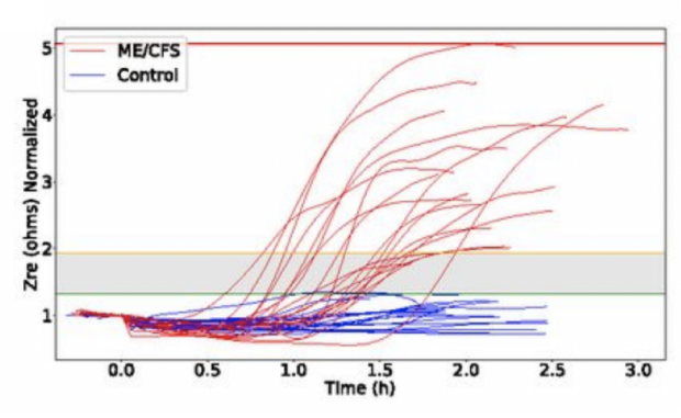
The first study investigated the electrophysiological properties of PBMCs in myalgic encephalomyelitis/chronic fatigue syndrome (ME/CFS); a debilitating disease of unknown pathophysiology with no reliable, validated, and quantitative diagnostic test. The dielectric and ζ-potential response of PBMCs to 1.5-hour hyperosmotic challenge differentiated ME/CFS donors from healthy controls with 81.80% sensitivity and 85.70% specificity. This shows potential as a quantitative diagnostic biomarker.
The second study examined whether the electrophysiological properties of PBMCs could act as a correlate of protection to SARS-CoV-2. Cytoplasmic conductivity in unchallenged PBMCs was significantly reduced in donors who had received three SARS-CoV-2 vaccine doses compared with unmatched COVID-19 naïve donors. Stimulation with the receptor binding domain of the SARS-CoV-2 spike protein resulted in significant differences in normalised values of membrane conductance in third-dose vaccinated donors, from COVID-19 naïve and second-dose donors.
The third study investigated chondrocytes, which are used extensively in cell-based cartilage-repair therapies. Chondrocytes rapidly dedifferentiate and become fibroblastic during monolayer cell culture – decreasing the success of reimplantation surgery. Significant changes in chondrocyte electrophysiological properties were observed over time in culture, laying the foundations for the identification of an electrophysiological biomarker that correlates with chondrocytic phenotype, to improve re-implantation outcome.
These studies demonstrate novel applications of dielectrophoresis and ζ-potential analysis – to quantitatively diagnose ME/CFS, identify changes in PBMCs following COVID-exposure, and changes in chondrocyte electrophysiology during dedifferentiation.
Files and links (1)
PDF
Krista Clarke Final Thesis 11.28 MB
PDF Embargoed Access, Embargo ends: 01/07/2025
Doctoral Thesis
Characterising the Electrophysiological Properties of Cells in Health and Disease
Krista Samantha Pauline Clarke
University of Surrey
Doctor of Philosophy (PhD), University of Surrey
28/06/2024
DOI:
https://doi.org/10.15126/thesis.901143
Abstract
Dielectrophoresis Myalgic Encephalomyelitis/ Chronic Fatigue Syndrome SARS-CoV-2 PBMC COVID-19 Chondrocyte Diagnosis
Biological cells possess intrinsic electrophysiological properties which are fundamental to cellular function. Changes in cell electrophysiology can act as a biomarker, for example to indicate transition from healthy to diseased cell states, changes in cell function, or cell differentiation. This thesis presents three studies which used dielectrophoresis (DEPtech 3DEP) and ζ-potential analysis (two fast, label-free, high-throughput, non-invasive, and low-cost tools) to examine the electrophysiological properties of two cell types, peripheral blood mononuclear cells (PBMCs) and chondrocytes, for novel medical applications.
The first study investigated the electrophysiological properties of PBMCs in myalgic encephalomyelitis/chronic fatigue syndrome (ME/CFS); a debilitating disease of unknown pathophysiology with no reliable, validated, and quantitative diagnostic test. The dielectric and ζ-potential response of PBMCs to 1.5-hour hyperosmotic challenge differentiated ME/CFS donors from healthy controls with 81.80% sensitivity and 85.70% specificity. This shows potential as a quantitative diagnostic biomarker.
The second study examined whether the electrophysiological properties of PBMCs could act as a correlate of protection to SARS-CoV-2. Cytoplasmic conductivity in unchallenged PBMCs was significantly reduced in donors who had received three SARS-CoV-2 vaccine doses compared with unmatched COVID-19 naïve donors. Stimulation with the receptor binding domain of the SARS-CoV-2 spike protein resulted in significant differences in normalised values of membrane conductance in third-dose vaccinated donors, from COVID-19 naïve and second-dose donors.
The third study investigated chondrocytes, which are used extensively in cell-based cartilage-repair therapies. Chondrocytes rapidly dedifferentiate and become fibroblastic during monolayer cell culture – decreasing the success of reimplantation surgery. Significant changes in chondrocyte electrophysiological properties were observed over time in culture, laying the foundations for the identification of an electrophysiological biomarker that correlates with chondrocytic phenotype, to improve re-implantation outcome.
These studies demonstrate novel applications of dielectrophoresis and ζ-potential analysis – to quantitatively diagnose ME/CFS, identify changes in PBMCs following COVID-exposure, and changes in chondrocyte electrophysiology during dedifferentiation.
Files and links (1)
Krista Clarke Final Thesis 11.28 MB
PDF Embargoed Access, Embargo ends: 01/07/2025
Last edited:


")
