Chronic CXCL10 alters neuronal properties in rat hippocampal culture, 2009, Cho et al

jnmaciuch

Senior Member (Voting Rights)
Chronic CXCL10 alters neuronal properties in rat hippocampal culture

Cho, Jungsook; Nelson, Thomas E.; Bajova, Hilda; Gruol, Donna L.

Abstract
The chemokine CXCL10 is expressed in the central nervous system (CNS) during neuroinflammatory conditions. Neurons express CXCR3, the receptor for CXCL10, and neuronal function has been shown to be altered by acute exposure to CXCL10. Little is known about the effects of chronic exposure to CXCL10 on neuronal function. Results from our studies show that chronic exposure of cultured rat hippocampal neurons to CXCL10 results in altered levels of protein for GABA and glutamate receptors and altered synaptic network activity. These effects of CXCL10 may contribute to altered CNS function that occurs in some chronic neuroinflammatory conditions.

Web | Journal of Neuroimmunology
https://doi.org/10.1016/j.jneuroim.2008.12.007 (Paywalled)
 
CXCL10 activation of CXCR3 receptors in neurons was found to be a driver of sickness behavior in animal models from this paper:

I thought this paper was interesting because if CXCL10 is relevant in ME/CFS, the findings from this paper could help explain some aspects of the illness that members have reported, specifically sensory sensitivity and finding relief from medications like Xanax that modulate neuron excitability.

In this thread I was also discussing the possibility of calcium flux in neurons causing mtDNA release under the right conditions, which would generate an interferon response in neurons:

This paper might provide an explanation for how that feedback loop could get locked in. With certain genetic predispositions, the feedback mechanism would be: Prolonged CXCL10 signaling from infection -> increased neuron excitability and therefore calcium flux -> increased mtDNA release through mitochondrial calcium channel VDAC -> mtDNA detecting by cGAS-STING triggers type I interferon -> type I interferon binds to IFNAR on brain epithelial cells, generating more CXCL10 -> [edit: prolonged CXCL10 signaling without infection, just driven by neuron firing].

Will come back to do more of a deep dive into the paper when I have a free moment. From my first read, the biggest limitation is that the findings come from cultured neurons, so may be missing an important physiological feedback mechanism that normally counteracts this effect in-vivo.
 
Last edited:
Overview of the paper:

Note: I don't have expertise in these specific methods, so am just assessing the general logic.

This is a brief paper on cultured neurons + glial cells from the rat hippocampus. Previous studies have shown differences in neuron function with acute CXCL10 exposure, but this study assessed the effects of prolonged exposure (9 days, according to the methods). Unless specified otherwise, each measurement had 6 technical replicates.

From Fig 1:
1762969555101.png
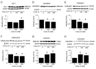
CXCL10 administration increased the expression of proteins involved in glutaminergic signaling and presynaptic neurotransmitter release (SYN1, NMDAR1, mGluR2/3) and decreased the expression of proteins involved in "inhibitory" GABA signaling.

From Fig 2:

1763063117637.png
To see if those differences in protein levels had an effect on spontaneous synaptic activity, they used a method that measures intracellular calcium oscillation. B is recordings in physiological saline, C is in the low Mg2+ condition. This is what the paper says about the low Mg2+ saline:
The reduced Mg2+ saline induces conditions reflective of strong synaptic drive by removing Mg2+ block of NMDARs.
I don't have the expertise to know whether this adequately mimics biological conditions, or if the apparent messiness of the oscillation patterns is to be expected. If the methodology makes sense, then this seems to suggest that chronic exposure to CXCL10 causes neurons to be more excitable in conditions that model more synaptic input.

Skipping to the last figure (Fig 4):

1763063695325.png
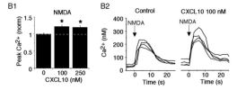
They tested the calcium oscillations in response to NMDA administration, and the CXCL10-exposed neurons showed higher total flux. I don't know what to make of the fact that the 250 nM CXCL10 condition was not higher than the 100--it might reflect a plateau of the effect of CXCL10 on NMDA-induced synaptic activity.

As already noted, the fact that these neurons were studied in-vitro is a major limitation, especially in the context of neuron excitability where input from other inhibitory networks or other physiological factors might play a large mitigating role. The findings from this specific paper are pretty limited, though they should be viewed in light of other papers which have shown similar effects of CXCL10.
 
This paper might provide an explanation for how that feedback loop could get locked in.
Nice loop. Could this also end up being in a sort of background low level state and ramped up state too do you think? I’m really interested in potential shifts in GABA and glutamate receptors over time and in response to activity/medications or immune activity.

Thanks for sharing the ideas and papers!
 
Prolonged CXCL10 signaling from infection -> increased neuron excitability and therefore calcium flux -> increased mtDNA release through mitochondrial calcium channel VDAC -> mtDNA detecting by cGAS-STING triggers type I interferon -> type I interferon binds to IFNAR on brain epithelial cells, generating more CXCL10 -> [edit: prolonged CXCL10 signaling without infection, just driven by neuron firing].

Loops like this do seem plausible. My caveats would be that if this loop is the normal rules then it should happen to all of us and if it only applies with a genetic nudge we want to know what the nudge is. It looks as if a genetic nudge may only provide a 10-20% lifetime chance of this happening, when we all get infections regularly. It also seems that this can only be operating in a very small part of the brain, since most of it works OK in ME/CFS. My main worry would be that randomly extracted hippocampal cells are very unlikely to be the relevant ones. The broader worry is that there are 101 loops like this that we could construct if we allow for them only to operate with a genetic tweak.

Nonetheless, sifting through candidate loops must be worth doing.

I am getting interested in the idea that we may be dealing with very local events specific to cell groups. The pain genetics people have identified about 20 separate cell populations in dorsal root ganglia, each doing different jobs - mechanical pain, proprioception, touch, etc. Each cell group expresses specific gene products. Absence of pain can occur with a gene defect affecting just one population.

An interesting implication of events in DRG, though, is that local loops ought to be independent. Feedback cycles in each of fifty DRG would not necessarily interact. So you might expect to get something more regional - which is of course what you see with "sudeck's atrophy" aka reflex sympathetic dystrophy affecting a hand or a foot. In the brain a local loop around cells in a basal nucleus like suprachiasmatic (where CLOCK is expressed) might work better.
 
The connection I still can't visualise is that between effects in the brain and the immune-type symptoms of ME/CFS.

I have swollen glands and a runny nose today after packing away a supermarket delivery yesterday. Standard stuff. But how do those tissues get the message that they should swell up and start being annoying? Might it be a different route to if (or when, more likely) I get the 'flu virus every third person seems to have at the moment?
 
I have swollen glands and a runny nose today after packing away a supermarket delivery yesterday. Standard stuff. But how do those tissues get the message that they should swell up and start being annoying? Might it be a different route to if (or when, more likely) I get the 'flu virus every third person seems to have at the moment?

I think that is a key argument. If the problem long-term is just in the brain then the brain must be sending out signals to lymphoid tissue to overdo things. While we know there are pathways that could allow that there is a spooky lack of evidence for it really happening in any known clinical context. Why is there no known brain disease that suddenly gives you swollen glands?

To me this is an encouraging part of the clinical story. It suggests that long-term persistence requires eom long-term overdoing within the immune system itself. Which of course is present in psoriasis and the like. And that sort of overdoing might be susceptible to daratumumab or something.

That still leaves room for brain loops being part of the problem at least for some people. Maybe the very severe people who cannot tolerate any stimuli have a brain loop on top of immune overdoing. Maybe for some the immune overdoing really has faded out and the brain is the problem. This was very much the question that seemed to be in Chris Ponting's mind when he first presented the DecodeME data. Do we have two stories or one or maybe most likely two modules to a story that can be mostly one or the other?
 
The connection I still can't visualise is that between effects in the brain and the immune-type symptoms of ME/CFS.

I have swollen glands and a runny nose today after packing away a supermarket delivery yesterday. Standard stuff. But how do those tissues get the message that they should swell up and start being annoying? Might it be a different route to if (or when, more likely) I get the 'flu virus every third person seems to have at the moment?
I posted this paper a while ago showing that interferon alone is enough to cause mucus production that is normally seen in response to respiratory viruses: https://www.s4me.info/threads/mucus...ers-hypoxia-of-covid-19-liu-et-al-2020.44922/

Haven’t seen specific research about swollen glands. But the main part of my theory that I’ve talked about across various threads is a transient interferon response caused by muscle use or neuron firing beyond a certain threshold for mtDNA release. Since we know that interferon alone can cause many of the symptoms associated with flu, the explanation is that a local reaction in one part of the body leaks a high enough level of interferon into the blood to reach other parts. That leakage is what normally happens in infections in the respiratory tract, when you get symptoms from a vaccine, flesh wounds causing sepsis, etc.
 
That still leaves room for brain loops being part of the problem at least for some people. Maybe the very severe people who cannot tolerate any stimuli have a brain loop on top of immune overdoing. Maybe for some the immune overdoing really has faded out and the brain is the problem. This was very much the question that seemed to be in Chris Ponting's mind when he first presented the DecodeME data. Do we have two stories or one or maybe most likely two modules to a story that can be mostly one or the other?
Is this similar to what Fluge, Mella and Tronstad suggested a while back?
 
I posted this paper a while ago showing that interferon alone is enough to cause mucus production that is normally seen in response to respiratory viruses: https://www.s4me.info/threads/mucus...ers-hypoxia-of-covid-19-liu-et-al-2020.44922/

Haven’t seen specific research about swollen glands. But the main part of my theory that I’ve talked about across various threads is a transient interferon response caused by muscle use or neuron firing beyond a certain threshold for mtDNA release. Since we know that interferon alone can cause many of the symptoms associated with flu, the explanation is that a local reaction in one part of the body leaks a high enough level of interferon into the blood to reach other parts. That leakage is what normally happens in infections in the respiratory tract, when you get symptoms from a vaccine, flesh wounds causing sepsis, etc.

I’m curious about how this theory explains the gradual baseline decline some people report after repeated PEM episodes. I think you posted about the mtDNA theory in other threads that I can't find at the moment.
 
I’m curious about how this theory explains the gradual baseline decline some people report after repeated PEM episodes
We don’t know if it is causal. I’d certainly not argue against be being so, as it did feel like that to me in some ways. But equally my decline could be explained by a general unrelated decline with those PEM periods as humps within it. So I’m not sure if’s a must have in any explanation.

Probably a bit like the background state and ramping up I mentioned. Not necessarily essential but I can imagine that if things got to a certain point they could self perpetuate there and no be able to be shift back down naturally maybe?
 
I’m curious about how this theory explains the gradual baseline decline some people report after repeated PEM episodes. I think you posted about the mtDNA theory in other threads that I can't find at the moment.
Yes, sorry, it is a bit scattered across threads at the moment. Since the theory itself is mostly pulling at threads as far as they can go from disparate observations, my attempt to explain some more specific parts of ME/CFS (beyond what aligns with observed phenomena, like the neuron excitability here or mucus production) would be hand-wavy at best. That’s largely why I’ve been more concerned with trying to get resources to prove or disprove any of my ideas rather than trying to shore everything up into a hypothesis paper.

If you’ll allow a bit of the hand-waving, my best guess is something to do with how repeated stimulation of the pathway in PEM keeps certain regions of the genome epigenetically “open” where they would otherwise stochastically get closed over time, which then gets locked in by the very same loop if the genes in those “open” regions are some of the ones that affect calcium metabolism and mtDNA release [Edit: like CD38]
 
Last edited:
I have not read through the original Fluge and Mella paper but I have heard of them talking about brain loops and synaptic changes much if at all. They seemed to be focusing on immune mechanisms?
I thunk they talk about various compensatory changes brought on by immune mechanisms, and that in some cases the compensatory changes (including neuronal ones) might persist even though the immune mechanisms have returned to a more normal state.
We suggest three principal steps underlie the initiation and maintenance of ME/CFS. (i) Immune response after infection serves as a triggering event, with a role for B cells/plasma cells and autoantibodies in the underlying pathology. (ii) The vascular system and possibly GPCRs are potential targets for autoantibodies, which may affect endothelium or neurovascular control and autonomic small nerve fibers. The autoantibodies could be pathogenic IgGs or functional autoantibodies that normally occur after infection, but persist and fail to resolve over time. This disturbed homeostasis involves endothelial dysfunction in large and small arteries, impaired venous return and preload failure, and arteriovenous shunting, presumed to result in impaired autoregulation of blood flow and tissue hypoxia on exertion. (iii) Secondary compensatory efforts may add to the clinical presentation and symptoms. They include autonomic adaptations, often with increased sympathetic tone, and metabolic adaptations aiming to restore energy supply.
In our model, clinical symptoms of ME/CFS are related primarily to the inadequate autoregulation of blood flow yielding tissue hypoxia on exertion, but are also influenced by the compensatory adaptations from increased sympathetic output and from metabolic shifts. We speculate that cognitive techniques, which are reported to help subgroups of patients, might act by modulating the sympathetic output. If so, one would expect a greater benefit for patients with less ongoing immune activation and less vascular dysregulation, but with main symptom contributions from the secondary autonomic adaptations. Conversely, patients with active immune disturbance and ongoing vascular dysregulation as the main symptom generators would have less impact from cognitive intervention, although psychosocial support and coping strategies may still have a beneficial impact on their quality of life.
 
I thunk they talk about various compensatory changes brought on by immune mechanisms, and that in some cases the compensatory changes (including neuronal ones) might persist even though the immune mechanisms have returned to a more normal state.

Yes, I don't think they were thinking of autonomous feedback loops in brain involving shifts in signalling thresholds. That is what the neuro-developmental and synapse-linked genes raise.
 
Back
Top Bottom