Community Symposium on the Molecular Basis of ME/CFS Sept 5 (Stanford/Ron Davis)

Ah, yes, I came back to the thread via an alert and missed that.

These are very interesting images. The 'blobby' nature of the signal is strongly suggestive of lymph node. It does not correspond to specific muscles or muscle groups and isn't really showing up in spine any more than the normal pattern. They have probably chosen a patient with a strong signal so everything is a bit brighter so the differential patterns are what matter.

The shoulder signal could be supraclavicular and axillary nodes and the pelvic signal inguinal and iliac nodes. Nevertheless, there is an odd skewing of signal spreading distally in both areas for a short distance, perhaps indicating uptake in draining lymphatics as they arrive at the major node sites. You can also get strange appearances a bit like this in conditions where lymph nodes are chronically enlarged and with cell activity secondary to distal disease like psoriasis - so-called dermatopathic lymphadenopathy. Maybe this could be a pattern of myopathic lymphadenopathy.

The images in polymyalgia are much more evenly diffuse and linear around muscles and joint capsules in should and pelvis so I don't think this is the same thing.
 
I have to say that this looks to me remarkably like exactly what we should have been looking for and expected. It suggests that immune cells are involved in certain activities within lymphoid organs, not within tissues normally targeted by inflammation, like skin, muscle, joint. Within a lymphoid organ you don't really get inflammation, except when there is breakdown of tissue architecture and pus or caseation as in a staph or TB lymphadenitis. Lymph node and spleen normally allow white cells to pass through freely and for spleen there is no real endothelial barrier at all so permeability does not change in the way it does in other tissues.

So the puzzle of there seeming to be abnormal ongoing immune cell activity without any inflammation or peripheral tissue pathology can be explained the obvious way - the activity is largely confined to lymphoid tissue itself. Beyond a certain distance from the trunk, muscle uptake looks quite normal but at least in this one image (edit: these two) node uptake is grossly abnormal.

This should please those who don't like brain theories!

I am surprised that, if this is a replicable finding, which I rather suspect it will be, nothing like this has been shown using, for instance, gallium scans. They show up in sarcoid nicely. But maybe this is a different sort of activity - (as Robert Phair and I have touched on).

Maybe this is some CD38 dependent cellular activity perhaps involving T/NK lymphocytes and macrophages/dendritic cells that responds to daratumumab?

So much of what we have seen in ME/CFS research doesn't replicate so one is always very cautious. However, quite unlike the Nakatomi pictures this looks to me like something specific and very interesting. It may turn out to be something else in people misdiagnosed but whatever it is it looks intriguing.
 
Last edited:
Before seeing these pictures I was getting around to posting a new thread on ME/CFS as "Host versus host disease" after a long discussion with Jo Cambridge, who has had graft versus host disease with her treatments. These pictures would be perfect for that. But it needs a separate thread.
 
Can’t find an email for Michelle James but she is on LinkedIn if anyone is on there and can contact her and invite her to the forum! Would be great if @Jonathan Edwards and Jo could connect with her. I know she has said too that she can design custom PET tracers and wanted to collaborate with anyone who had ideas.

 
This is really exciting stuff! Do we have any information on the severity of the patients who were scanned? It would be interesting to see if the picture was even clearer in severe/very severe patients if they have scanned mild/moderate patients here.
 
Not sure if we have images for the GPR84 receptor?
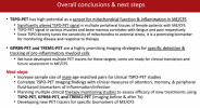
No, I don't think they have tried in ME/CFS patients yet (maybe not even any humans). She talks about how they are developing the GPR84 tracers and gives rationale for using them (more specific to microglia and macrophages than TSPO). She said they're at the stage where they're about to translate them to clinical use.

She also says they're interested for looking at the TREM1 marker in ME/CFS patients (more specific to peripheral immune cells like monocytes, macrophages, and neutrophils), but that so far it appears to be useful for MS imaging.

The summary of all of it:
1757161940135.png
 
This is really exciting stuff! Do we have any information on the severity of the patients who were scanned? It would be interesting to see if the picture was even clearer in severe/very severe patients if they have scanned mild/moderate patients here.
I don't know about the two specific people in the images, but in the demographic data screenshot for all 13 women, for the multidimensional fatigue inventory (MFI) score it says 86.62 for cases and 32.08 for controls.

From a paper:
A priori, cases were classified into tertiles for ME/CFS severity: MFI-20 scores from 51–75 were classified as mild disease, scores from 76–85 as moderate disease, and scores from 86–100 as severe disease.
 
So the puzzle of there seeming to be abnormal ongoing immune cell activity without any inflammation or peripheral tissue pathology can be explained the obvious way - the activity is largely confined to lymphoid tissue itself.
In the screenshot I posted of the bar plots of the data for tissues in everyone, it looks like there may be a signal in the bone as well. In the pelvic bone and maybe also in the femur and lumbar bone marrow, though not significant for the last two.

Does it change things for there to be signal in bone as well as lymph nodes?
 
Back
Top Bottom