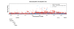
Preprint Initial findings from the DecodeME genome-wide association study of myalgic encephalomyelitis/chronic fatigue syndrome, 2025, DecodeMe Collaboration

Genetic links to "intelligence" always struck me as a hollow concept anyways. Really, it's a genetic association for doing slightly better on a handful of tests where you match patterns or pick words out of a list. The links between those types of tests and anything else people would associate with "intelligence"--good decision making, creative problem solving, professional success, interest in research, etc.--have always come across as incredibly dubious to me. Not in the least because social factors so heavily skew both performance on those tests and any of those other indicators of "intelligence". Is a gene actually associated with the nebulous concept of "intelligence", or with the closed-off social strata that have better access to schooling and more time on their hands to participate in research, or perhaps with the lack of various health conditions that would make someone less focused during a long battery of cognitive tests?

There may well be some confounding and self-selection with that particular finding, but more likely explained by those other factors rather than any concept of "intelligence."
Not to mention the cultural bias in the questions themselves having blown a lot of the old tests as not measuring what they thought they were (and then it’s so obvious you can’t unsee it after it’s pointed out) in the last decade.
 
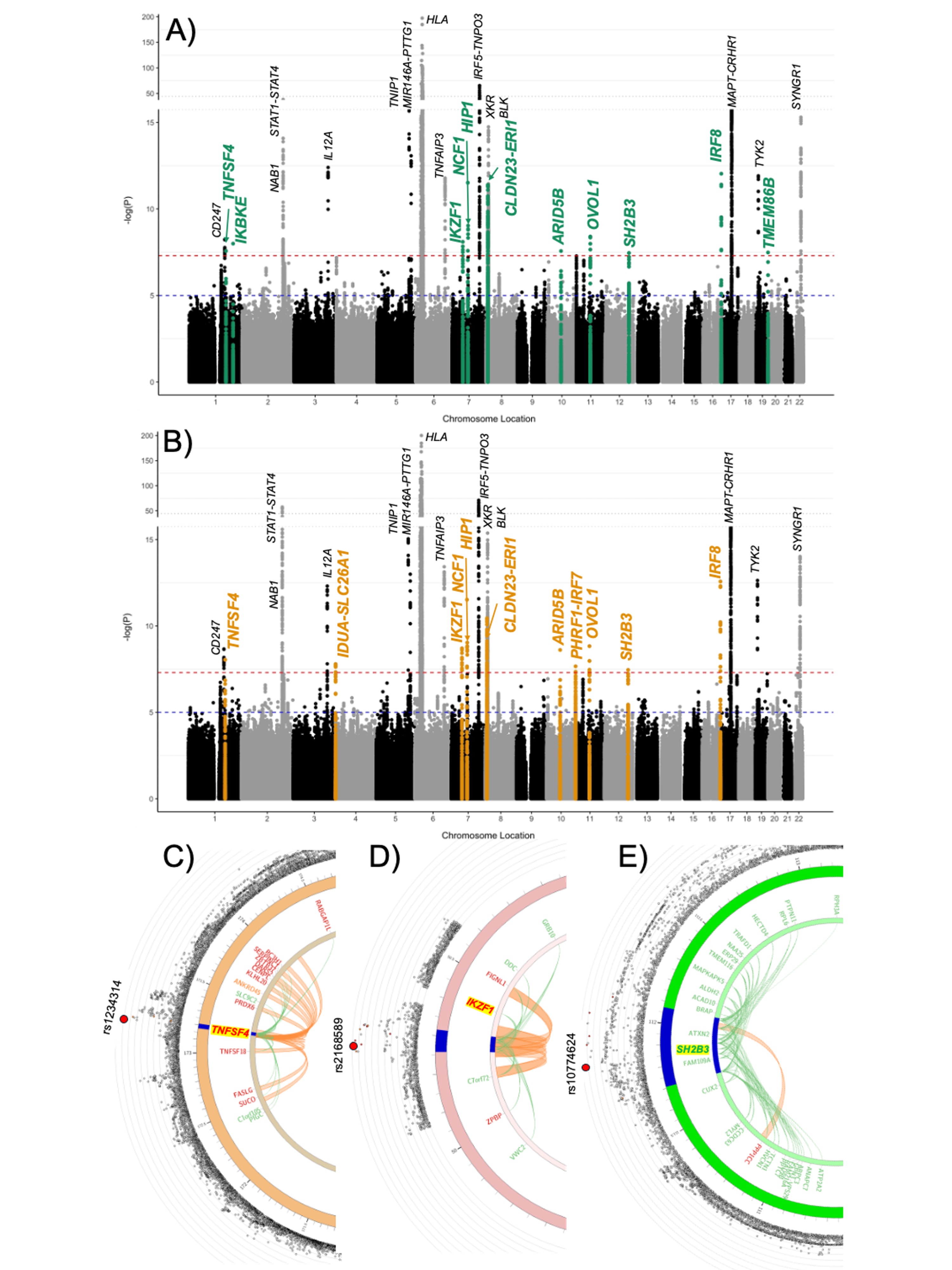
There's also this blog by Paolo Maccalini on the DecodeME results, focusing on the FUMA SNP2GENE analysis, which forestglip explored earlier in this thread.

The 18 genes singled out are mostly associated with the nervous system (both CNS and associated with peripheral nerve disease, injury or recovery), the immune system and mitochondria.

eg starting with the first one, ABT1 is a mitochondrial associated gene but is associated with the IGHMBP2 related genetic motor neuron diseases.
 
Last edited:
The 18 genes singled out are mostly associated with the nervous system (both CNS and associated with peripheral nerve disease, injury or recovery), the immune system and mitochondria.

eg starting with the first one, ABT1 is a mitochondrial gene but is associated with the IGHMBP2 related genetic motor neuron diseases.
I have nowhere close to enough time to look yet... is this mitochondrially encoded genes or mitochondrially localised gene products? Or just with some association with mitochondrially relevant pathways?
 
On our blog, Paolo wrote:
My fine-mapping attempt of DecodeME was performed using SusieR with Linkage Disequilibrium matrices from the original UK Biobank (downloaded from the Broad Institute repository). In order to use them, I had to lift over the DecodeME summary statistics from GRCh38 to GRCh37. This is not a perfect approach because there is a loss of about half of the variants. But it is the best I could do...
This is curious. When using the GenomicRanges and rtracklayer packages in R we only lost about 25.000 variants out of almost 9 million. FUMA/MAGMA report the same in the log file: “25262 positions did not align with the GRCh37 reference.”
 
Previous work has shown that men with Klinefleter’s syndrome (47,XXY) as well as women with 47,XXX are found in excess among SLE patients well as among Sjogren’s disease, systemic sclerosis and idiopathic inflammatory myositis.
This is really interesting. Looking at the number of patients that have an extra X chromosome helped nail down that the X chromosome was a risk factor for SLE. Here are the two papers it cites for the above:

Klinefelter's syndrome (47,XXY) in male systemic lupus erythematosus patients: Support for the notion of a gene-dose effect from the X chromosome (2008)
Objective
Systemic lupus erythematosus (SLE) is a systemic autoimmune disease that predominantly affects women. Despite isolated reports of patients with coexisting Klinefelter's syndrome (47,XXY) and SLE, no association of Klinefelter's syndrome with SLE or any other autoimmune disease has been established. The present study was undertaken to investigate the prevalence of Klinefelter's syndrome in a large population of patients with SLE.

Methods
Sex chromosome genotyping was performed in 981 SLE patients, of whom 213 were men. A first group of 844 SLE patients from 378 multiplex families and a second group of 137 men with nonfamilial SLE were evaluated. In selected cases, chromosomes were enumerated by fluorescence in situ hybridization (FISH) and karyotyping in transformed B cell lines.

Results
Of 213 men with SLE, 5 had Klinefelter's syndrome (1 in 43). Four of them were heterozygous at X markers, and Klinefelter's syndrome was confirmed by FISH and karyotyping in the fifth. An overall rate of 47,XXY of 235 per 10,000 male SLE patients was found (95% confidence interval 77–539), a dramatic increase over the known prevalence of Klinefelter's syndrome in an unselected population (17 per 10,000 live male births). Asking men with SLE about fertility was highly sensitive (100%) for Klinefelter's syndrome. All 768 women with SLE were heterozygous at X.

Conclusion
The frequency of Klinefelter's syndrome (47,XXY), often subclinical, is increased in men with SLE by ∼14-fold compared with its prevalence in men without SLE. Diagnostic vigilance for 47,XXY in male patients with SLE is warranted. These data are the first to show an association of Klinefelter's syndrome with an autoimmune disease found predominantly in women. The risk of SLE in men with Klinefelter's syndrome is predicted to be similar to the risk in normal women with 46,XX and ∼14-fold higher than in men with 46,XY, consistent with the notion that SLE susceptibility is partly explained by an X chromosome gene-dose effect.
Web | Arthritis and Rheumatology | Paywall

X Chromosome Dose and Sex Bias in Autoimmune Diseases: Increased Prevalence of 47,XXX in Systemic Lupus Erythematosus and Sjögren's Syndrome (2015)
Objective
More than 80% of autoimmune disease predominantly affects females, but the mechanism for this female bias is poorly understood. We suspected that an X chromosome dose effect accounts for this, and we undertook this study to test our hypothesis that trisomy X (47,XXX; occurring in ∼1 in 1,000 live female births) would be increased in patients with female-predominant diseases (systemic lupus erythematosus [SLE], primary Sjögren's syndrome [SS], primary biliary cirrhosis, and rheumatoid arthritis [RA]) compared to patients with diseases without female predominance (sarcoidosis) and compared to controls.

Methods
All subjects in this study were female. We identified subjects with 47,XXX using aggregate data from single-nucleotide polymorphism arrays, and, when possible, we confirmed the presence of 47,XXX using fluorescence in situ hybridization or quantitative polymerase chain reaction.

Results
We found 47,XXX in 7 of 2,826 SLE patients and in 3 of 1,033 SS patients, but in only 2 of 7,074 controls (odds ratio in the SLE and primary SS groups 8.78 [95% confidence interval 1.67–86.79], P = 0.003 and odds ratio 10.29 [95% confidence interval 1.18–123.47], P = 0.02, respectively). One in 404 women with SLE and 1 in 344 women with SS had 47,XXX. There was an excess of 47,XXX among SLE and SS patients.

Conclusion
The estimated prevalence of SLE and SS in women with 47,XXX was ∼2.5 and ∼2.9 times higher, respectively, than that in women with 46,XX and ∼25 and ∼41 times higher, respectively, than that in men with 46,XY. No statistically significant increase of 47,XXX was observed in other female-biased diseases (primary biliary cirrhosis or RA), supporting the idea of multiple pathways to sex bias in autoimmunity.
Web | American College of Rheumatology | Paywall

Notably, they were able to find an excess of SLE patients that have extra X chromosomes with only 213 males and 2826 females. DecodeME has far more cases than this, so if the X chromosome is similarly involved in ME/CFS, maybe people with extra X chromosomes would be over-represented in the cohort.

I wonder if DecodeME is equipped to analyze this. @Chris Ponting maybe you could let us know if this is something you plan to look at?
 
When I previously ran LDSC to test for genetic correlations between DecodeME and all the traits in the UK Biobank, the correlation with lupus didn't work, likely because the sample size in the Biobank was too small.

Recent discussions about lupus made me want to see if I could get it working with a different dataset. I found a larger SLE study, Bentham 2015 [1], and the summary statistics for this study are downloadable from a couple locations, like Open GWAS. This website says the study includes 5201 cases and 9066 controls*.

I ran LDSC between the DecodeME data and the SLE data, again using Bigagwas, and here are the results:
Heritability of phenotype 1 [ME/CFS]
---------------------------
Total Liability scale h2: 0.0885 (0.0058)
Lambda GC: 1.1019
Mean Chi^2: 1.1428
Intercept: 0.9089 (0.0075)
Ratio < 0 (usually indicates GC correction).

Heritability of phenotype 2/2 [Systemic lupus erythematosus]
-----------------------------
Total Liability scale h2: 0.1789 (0.022)
Lambda GC: 1.4673
Mean Chi^2: 1.3551
Intercept: 1.2021 (0.0093)
Ratio: 0.5692 (0.0262)

Genetic Covariance
------------------
Total Liability scale gencov: 0.0334 (0.0071)
Mean z1*z2: 0.0341
Intercept: -0.0212 (0.0062)

Genetic Correlation
-------------------
Genetic Correlation: 0.2658 (0.0535)
Z-score: 4.9637
P: 6.9154e-07
So genetic correlation is 0.27, which is small but not nothing, and the correlation is significant at p=6.9e-7.

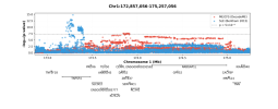
I thought it might be interesting to look at some of the significant regions to see how they compare between the two studies.

Here is that RABGAP1L region. SLE is in blue and ME/CFS is in red.
decodeme-sle_chr1:172857056-175257056.png

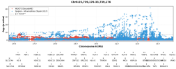
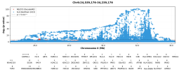
chr6p22.2:
decodeme-sle_chr6:25739176-26739176.png

Not much else is very significant for SLE around the regions of the rest of the top 8 ME/CFS loci.

I think the numbers given for sample size on Open GWAS might be incorrect. When looking at the paper, the numbers given match with the sample size for the meta-analysis which included the main GWAS plus the Hom et al data.

But the p-values in the summary statistics seem to match the results from only the main GWAS when looking at Supplementary Table 3a. This would mean the sample size would be 4036 SLE cases and 6959 controls.

I used the larger sample size given by Open GWAS for LDSC, but this might not give exactly the right results if it should actually be the smaller sample size, which is about 25% smaller.

1. Bentham, James et al. “Genetic association analyses implicate aberrant regulation of innate and adaptive immunity genes in the pathogenesis of systemic lupus erythematosus.” Nature genetics vol. 47,12 (2015): 1457-1464. doi:10.1038/ng.3434 https://pmc.ncbi.nlm.nih.gov/articles/PMC4668589/
 
The RABGAP1L patterns look mutually exclusive, which might be interesting?
There's a lot of genes in the area. It might be that the loci relate to totally different genes and their proximity is a coincidence.

Though DNA is a very, very long thing, so it kind of seems unlikely to me to happen to be right next to each other by chance. But possible.
 
The lupus locus on chromosome 1 above is right above TNFSF4 (AKA OX-40 ligand). There are a lot of papers talking about the connection between this gene and lupus, for example:

Polymorphism at the TNF superfamily gene TNFSF4 confers susceptibility to systemic lupus erythematosus (2013)
Systemic lupus erythematosus (SLE) is a multisystem complex autoimmune disease of uncertain etiology (OMIM 152700). Over recent years a genetic component to SLE susceptibility has been established1–3. Recent successes with association studies in SLE have identified genes including IRF5 (refs. 4,5) and FCGR3B6.

Two tumor necrosis factor (TNF) superfamily members located within intervals showing genetic linkage with SLE are TNFSF4 (also known as OX40L; 1q25), which is expressed on activated antigen-presenting cells (APCs)7,8 and vascular endothelial cells9, and also its unique receptor, TNFRSF4 (also known as OX40; 1p36), which is primarily expressed on activated CD4+ T cells10.

TNFSF4 produces a potent co-stimulatory signal for activated CD4+ T cells after engagement of TNFRSF4 (ref. 11).

Using both a family-based and a case-control study design, we show that the upstream region of TNFSF4 contains a single risk haplotype for SLE, which is correlated with increased expression of both cell-surface TNFSF4 and the TNFSF4 transcript.

We hypothesize that increased expression of TNFSF4 predisposes to SLE either by quantitatively augmenting T cell–APC interaction or by influencing the functional consequences of T cell activation via TNFRSF4.
Web | PDF | Nature Genetics | Open Access

Blockade of OX40/OX40L signaling using anti-OX40L alleviates murine lupus nephritis (2024)
Genetic variants of the OX40 ligand (OX40L) locus are associated with the risk of systemic lupus erythematosus (SLE), it is unclear how the OX40L blockade delays the lupus phenotype. Therefore, we examined the effects of an anti-OX40L antibody in MRL/Lpr mice. Next, we investigated the effect of anti-OX40L on immunosuppression in keyhole limpet hemocyanin-immunized C57BL/6J mice. In vitro treatment of anti-OX40L in CD4+ T and B220+ B cells was used to explore the role of OX40L in the pathogenesis of SLE.

Anti-OX40L alleviated murine lupus nephritis, accompanied by decreased production of anti-dsDNA and proteinuria, as well as lower frequencies of splenic T helper (Th) 1 and T-follicular helper cells (Tfh).

In keyhole limpet hemocyanin-immunized mice, decreased levels of immunoglobulins and plasmablasts were observed in the anti-OX40L group. Anti-OX40L reduced the number and area of germinal centers. Compared with the control IgG group, anti-OX40L downregulated CD4+ T-cell differentiation into Th1 and Tfh cells and upregulated CD4+ T-cell differentiation into regulatory T cells in vitro.

Furthermore, anti-OX40L inhibited toll-like receptor 7-mediated differentiation of antibody-secreting cells and antibody production through the regulation of the SPIB-BLIMP1-XBP1 axis in B cells.

These results suggest that OX40L is a promising therapeutic target for SLE.
Web | PDF | Eur J Immunol. | Open Access

Edit: Aha! It was mentioned in the DecodeME candidate genes document as a potential gene that this locus applies to (thanks Evergreen for posting about this):
TNFSF4 (Tier 1)

Protein: Tumor necrosis factor ligand superfamily member 4, also known as CD252 or OX-40L. UniProt. GeneCards. The allele that increases the risk of ME/CFS is associated with decreasing TNFSF4 gene expression.​
Molecular function: Cytokine that promotes T-cell proliferation, differentiation, and survival (12). Expressed on CD4+ T cells, CD8+ T cells and vascular endothelial cells.​
Cellular function: Regulates T-cell-mediated immunity.​
Link to disease: Region upstream of TNFSF4 is associated with systemic lupus erythematosus risk (13).​
Potential relevance to ME/CFS: Potential dysregulation of T-cell mediated immunity.​

The DecodeME paper says that the ME/CFS variants here are associated with decreased expression of TNFSF4 in the lung, skin of sun exposed lower leg, and thyroid.
 
Last edited:
Blockade of OX40/OX40L signaling using anti-OX40L alleviates murine lupus nephritis (2024)


A few OX40L monoclonals are coming to market soon:

"normalize the overactive immune system, without depleting T cells"

Originally for dermatitis, but I believe they think they will be good general immune T-cell suppression, possibly off-label
 
Thanks for this analysis @forestglip ! I think this is incredibly useful precisely because it's a comparison to what a GWAS would look like in an illness where we know TNF and BTNs are relevant in the pathophysiology (and can therefore be much more confident that the associations in the SLE cohort are driven by those genes).

It's interesting that you can see a similar indication of LD in the entire RABGAP1L region that we were discussing in another thread--the little island of blue pretty much confirms that the whole region tends to get inherited together. But there was an open question as to whether the little stretch right near DARS2 is being confounded by LD with RABGAP1L. I can't say this with complete confidence given the small cohort, but seeing the absence of an "LD island" around DARS2 in SLE makes me more confident that the relevant genes in that region for ME/CFS are DARS2 (or its immediately adjacent genes) and RABGAP1L separately (weakly).

Similarly, there was an open question of whether the hits on Chr6 were in relevant regulatory regions for BTNs. From the blue dots, we get a clear picture of exactly where the strongest regulatory regions are upstream of the BTNs, and it's not where the [edit: strongest] of the associated SNPs in ME/CFS are lying. And, even more interestingly, the region that is most highly associated with ME/CFS is almost entirely devoid of hits in the SLE cohort. Being cautious again about overinterpreting, I'd say this is one piece of evidence showing that it really is the histone proteins in that region that are relevant to ME/CFS.
 
Last edited:
Would it be worthwhile in trying to get those researchers who are already looking at the genetics of lupus (or other similar conditions) interested in the results of DecodeME?

Yes. The King's people are obviously interested. They also have an interest in pain/fibromyalgia genetics. I don't have contacts ith them but this should be discussed on Nov 6th.
 
Back
Top Bottom