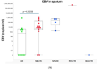
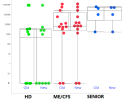
Now published. Minor changes to abstract wording. I haven't checked the differences in the full text. Though the inconsistency in the EBV chart is still there.
Prevalence of EBV, HHV6, HCMV, HAdV, SARS-CoV-2, and Autoantibodies to Type I Interferon in Sputum from Myalgic Encephalomyelitis/Chronic Fatigue Syndrome Patients
Ulf Hannestad, Annika Allard, Kent Nilsson, Anders Rosén
[Line breaks added]
Abstract
An exhausted antiviral immune response is observed in myalgic encephalomyelitis/chronic fatigue syndrome (ME/CFS) and post-SARS-CoV-2 syndrome, also termed long COVID. In this study, potential mechanisms behind this exhaustion were investigated.
First, the viral load of Epstein–Barr virus (EBV), human adenovirus (HAdV), human cytomegalovirus (HCMV), human herpesvirus 6 (HHV6), and severe acute respiratory syndrome coronavirus 2 (SARS-CoV-2) was determined in sputum samples (n = 29) derived from ME/CFS patients (n = 13), healthy controls (n = 10), elderly healthy controls (n = 4), and immunosuppressed controls (n = 2). Secondly, autoantibodies (autoAbs) to type I interferon (IFN-I) in sputum were analyzed to possibly explain impaired viral immunity.
We found that ME/CFS patients released EBV at a significantly higher level compared to controls (p = 0.0256). HHV6 was present in ~50% of all participants at the same level. HAdV was detected in two cases with immunosuppression and severe ME/CFS, respectively. HCMV and SARS-CoV-2 were found only in immunosuppressed controls.
Notably, anti-IFN-I autoAbs in ME/CFS and controls did not differ, except in a severe ME/CFS case showing an increased level.
We conclude that ME/CFS patients, compared to controls, have a significantly higher load of EBV. IFN-I autoAbs cannot explain IFN-I dysfunction, with the possible exception of severe cases, also reported in severe SARS-CoV-2. We forward that additional mechanisms, such as the viral evasion of IFN-I effect via the degradation of IFN-receptors, may be present in ME/CFS, which demands further studies.
Link | PDF (Viruses) [Open Access]


