Transcriptome analysis of classical blood cells reveals down-regulation of proinflammatory genes in the classical monocytes of LC patients,2025,Fricke

Chandelier

Senior Member (Voting Rights)

Full title: Transcriptome analysis of classical blood cells reveals down-regulation of proinflammatory genes in the classical monocytes of Long-COVID patients

Florian  FrickeFlorian FrickeFranz  MaiFranz Mai
Christine  Wossidlo
Christine Wossidlo
Felix  Steinbeck
Felix SteinbeckWendy  Bergmann-EwertWendy Bergmann-EwertMarcel  KordtMarcel KordtKarin  KraftKarin KraftBritta  MüllerBritta MüllerEmil  Christian ReisingerEmil Christian ReisingerBrigitte  Müller-HilkeBrigitte Müller-Hilke*

Abstract
Despite extensive research, the pathogenesis and predispositions underlying Long-COVID remain poorly understood.

To address this, we analyzed the immunological landscapes of 44 Long-COVID patients and 44 matched convalescents using single-cell RNA sequencing (scRNA-seq) of peripheral blood mononuclear cells (PBMCs) and validated findings with plasma cytokine measurements via Luminex technology.
While immune cell compositions showed minimal quantitative differences only among NK cells, transcriptome analyses identified distinct gene expression patterns, particularly in classical monocytes: Long-COVID patients exhibited downregulation of inflammation-associated genes, including IL1B and CXCL2.
Imputation of transcription factor activity hinted at reduced inflammasome activity (via SNAI1) and impaired monocyte differentiation (via ATF2) in Long-COVID. RNA velocity supported the presence of immature classical monocytes in patients.

These findings showed that monocytes might be dysregulated and/or exhausted in Long-COVID patients.
 
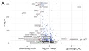
UMAP clustering of cells revealed distinct expression profiles between patients and controls in classical and intermediate, but not non-classical, monocytes. Differential gene expression analysis, based on a fold change threshold greater than 3 and a p-value threshold below 10−18, identified 40 DEGs in classical, 13 in intermediate, and 6 in non-classical monocytes, while other cell types showed minimal changes.

The volcano plot in Figure 4A details these findings for the classical monocytes. In total, there were 37 downregulated genes in patients with long COVID, among them IL1B, several chemokines (CCL3, CCL4, CXCL1, CXCL2, CXCL3, and CXCL8), and the TNFa-induced protein 3 (TNFAIP3). Further downregulated genes included DUSP2, encoding a phosphatase that downregulates members of the mitogen-activated protein (MAP) kinase superfamily, and MIR155HG, a microRNA involved in the regulation of MHCII antigen presentation.

Screenshot 2025-11-08 at 3.00.00 PM copy.jpg

Patients with long COVID showed a significantly reduced expression of the inflammation-related pathways, including TNFa signaling via NF-kB, consistently through all subsets of monocytes. Additional pathways, such as inflammatory response and apoptosis, were also downregulated, while heme metabolism and hallmark genes of oxidative phosphorylation (although failing to reach significance) demonstrated a higher NES in patients compared to the controls.

the transcriptional profile of classical monocytes implied not only a lower expression of pro-inflammatory cytokines and various chemokines but also an involvement of TFs that suppress inflammasome activity in patients. A conspicuous deficiency of transcriptional regulation promoting monocyte differentiation was observed, indicating an immature phenotype in the classical monocytes of patients.

we did not find any robust correlation between the gene expression profiles of patients and the clinical assessments, which may indicate that the distinct immunological changes in patients with long COVID contribute to rather diverse phenotypic disease characteristics.

In conclusion, we here present a downregulation of the inflammatory pathways in the classical monocytes of patients with long COVID.
 
I would like to think studies like this could tell us something. However, it seems likely that they are simply looking at how old the cells are or how much chemical noise they have encountered while circulating from the body moving about.

People talk of classical monocytes, which do not express much CD16. But if you put them in culture they express CD16 nicely. So CD16 is not a lineage marker.

Rather than talk of downregulation why isn't this just less upregulation? Circulating monocytes are small cells not doing much waiting to be called out into tissues if needed. Maybe if people are inactive the monocytes are not getting any signals they might be needed.
 
Back
Top Bottom