A causal link between autoantibodies and neurological symptoms in long COVID, 2026, Santos Guedes de Sa, Iwasaki et al

EndME

Senior Member (Voting Rights)
Now published - see here


A causal link between autoantibodies and neurological symptoms in long COVID

Abstract

Acute SARS-CoV-2 infection triggers the generation of diverse and functional autoantibodies (AABs), even after mild cases. Persistently elevated autoantibodies have been found in some individuals with long COVID (LC). Using a >21,000 human protein array, we identified diverse AAB targets in LC patients that correlated with their symptoms. Elevated AABs to proteins in the nervous system were found in LC patients with neurocognitive and neurological symptoms. Purified Immunoglobulin G (IgG) samples from these individuals reacted with human pons tissue and were cross-reactive with mouse sciatic nerves, spinal cord, and meninges. Antibody reactivity to sciatic nerves and meninges correlated with patient-reported headache and disorientation. Passive transfer of IgG from patients to mice led to increased sensitivity and pain, mirroring patient-reported symptoms. Similarly, mice injected with IgG showed loss of balance and coordination, reflecting donor-reported dizziness. Our findings suggest that targeting AABs could benefit some LC patients.

https://www.medrxiv.org/content/10.1101/2024.06.18.24309100v1?rss=1&utm_source=dlvr.it&utm_medium=twitter
 
Last edited by a moderator:
I would like to see how well controlled this study is.
I am a bit leery of antibodies that react both to meninges and sciatica nerves being related to headache. It would also be more convincing if they mentioned a bit about what they didn't find too.
 
It seems "causal" is used somewhat vaguely here.

The data on the cohort seems to be lacking. I can't see any information on illness duration, last known infection, BMI, comorbidities, etc. Furthermore whilst they started of with a large cohort in later experiments they are using various different subsets of this cohort and I can't see any details on these subsets or how these subsets were selected.

Other than that they did a lot of IgG transfer mice experiment. Hard to tell if anything is a signal there or rather noise and some results seem to contradict those of den Dunnen. For example I don't think there's much evidence to suggest that grip strength test tells us much in humans with neurological LC (there's some evidence by Scheibenbogen but also different results from various other studies for general LC or ME), I think such experiments on mice are even more speculative especially if grip strength is supposed to be correlated with tinnitus.
 
Last edited:
I think the pain aspect with impact on SFN is fascinating and important.

Really that's the stand out piece for me.

They took IgG from patients that experienced pain and put them in mice, which then created increased pain in the mice. They took a cohort from these mice to look at small fibers and find enhanced SFN in those that got the IgG from patients experiencing SFN. That's pretty convincing to me from first read.

We might be seeing IgG as an initiator of autonomic dysfunction. That's the potentially causal aspect I think they refer in the title.
 
They took a cohort from these mice to look at small fibers and find enhanced SFN in those that got the IgG from patients experiencing SFN. That's pretty convincing to me from first read.

Screen Shot 2024-06-20 at 6.44.31 pm.png
4 long covid; 4 healthy controls - a separate mouse for each person and (I think) the two sampling times.


Figure 4p
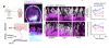
P Longitudinal Two-photon imaging of Scn10a-Cre::td Tomato reporter mice reveals collagen fibers by second harmonic generation (SHG) as well as nociceptor axons and the perpendicular intraepidermal nerve fibers (IENF; white/pink). Quantification of total IENF volume and number of fibers crossing the dermis boundary after passive transfer. Scale bars 50 m. Each dot in the figure represents the value obtained from an individual mouse. Each μ
mouse received antibodies isolated from a single human participant. Data are presented as the mean. Significant p values are described in the image, as determined by T-test for two groups comparison or one-way ANOVA corrected for multiple comparisons with Tukey test for multiple groups comparison.

We observed that mice injected with IgG from patients with LC had a reduction in the number and volume of Intraepidermal Nerves Fibers (IENF) when compared with mice that received IgG from healthy controls (Figure 4P). The reduction in the IENF is a marker for Small Fiber Neuropathy (SFN) 46 that usually presents with chronic pain described as burning pain and/or pins and needles sensations (both symptoms were associated with the hot plate test results) (Figure 4F-G). We also evaluated anxiety-like behavior and locomotion by open field and elevated zero maze (Supp. Fig. 6A-C), and blood pressure and heart rate (Supp. Fig. 6D) to evaluate pathological changes to orthostatic responses. We did not observe significant differences in these tests between the groups receiving IgG from HC, CC, or LC participants.
 
Last edited:
I'm quite taken by this paper. I like the way they've not just hunted for auto-antibodies but tried to see what effects those autoantibodies might have on tissues and whole organisms.

I don't think they're claiming definitive proof that AABs are the cause of long covid, but they do provide a solid link from some of the AABs to symptom load in some patients.

The happy thing about pointing the finger at AABs is we have a mechansim to attack them, via ivig/plasmapheresis. So the paper leads naturally to an interventional trial where they define a subset with high neurological symptom load (and/or high measured AABs) and see if they feel different after ivig. I'm very keen on adding definition to the subset of people ivig/plasmapheresis might help, since we know it does seem to help some.

The paper will make a splash since it's from Yale, and so it's pleasing to see it is fairly solid paper.
 
They took IgG from patients that experienced pain and put them in mice, which then created increased pain in the mice. They took a cohort from these mice to look at small fibers and find enhanced SFN in those that got the IgG from patients experiencing SFN. That's pretty convincing to me from first read.

4 long covid; 4 healthy controls - a separate mouse for each person and (I think) the two sampling times.

Have you ever done experiments on pain sensitivity in mice, Chris?
To me this is far too good ever to be true. And it doesn't really add up if you look at detail as ever.

To get a meaningful result with a system like this you would need a drug company unit running fresh experiments every fortnight for eighteen months to get the quality control you need.
 
Have you ever done experiments on pain sensitivity in mice, Chris?
To me this is far too good ever to be true. And it doesn't really add up if you look at detail as ever.

To get a meaningful result with a system like this you would need a drug company unit running fresh experiments every fortnight for eighteen months to get the quality control you need.

Can you give us an example for the kind of mistaken procedure that would lead to SFNP in 4 mice but not in the other 4? How would that happen?

Personally, I don't think ME/CFS is a B-cell mediated disease, but it might be for some, maybe the mistake that we keep on making is not to subgroup patients. Injecting ABs in mice (until we know more) could be a way to subgroup patients?

Additionally, is it not conceiveable that various (differing from patient A to patient B) ABs lead to the same or similar enough downstream pathology? Maybe our mistake has been thinking that we will find one AB that explains it all?

We already kind of know from the NIH intramural study that 40% of pwME are misdiagnosed, which means nothing other than 'we have a better explanation' for these patients. Maybe patients with ABs eliciting such responses in mice will be another group of patients for which there are better explanations, at least for parts of what they individually consider 'their version of ME'.

Not all ME/CFS paients have SFNP and suffer from pain; both aren't necessary for an ME/CFS diagnosis, they are real issues nevertheless, and they need solutions.

Thank you!
 
Last edited:
Can you give us an example for the kind of mistaken procedure that would lead to SFNP in 4 mice but not in the other 4? How would that happen?


Just that the method of measurement may be unreliable - for a hundred different reasons.

We have seen how consistent.y poor methodology is in the psychiatric sector. I am afraid to say that it tends to be almost as bad in this sort of biomedical area. Everyone wants to report a positive result and most of the time not enough standardisation has been done to ensure the experiments mean anything.

If there are problems with choosing the right patients it becomes even more improbable that you would get a significant result that is meaningful. Post hoc rationalisation of why experiments did not quite do what you expected are what bad science feeds off for ever.
 
Just that the method of measurement may be unreliable - for a hundred different reasons.

We have seen how consistent.y poor methodology is in the psychiatric sector. I am afraid to say that it tends to be almost as bad in this sort of biomedical area. Everyone wants to report a positive result and most of the time not enough standardisation has been done to ensure the experiments mean anything.

If there are problems with choosing the right patients it becomes even more improbable that you would get a significant result that is meaningful. Post hoc rationalisation of why experiments did not quite do what you expected are what bad science feeds off for ever.

The way forward then would be better standardisation.
 
Now published:

A causal link between autoantibodies and neurological symptoms in long COVID

Santos Guedes de Sá, Keyla; Silva, Julio; Bayarri-Olmos, Rafael; Baker, Christopher A.; Lu, Zhenni; Gipson, Wilson; Na, Daxiang; Chen, Bandy; Wenxue, Li; Khosroabadi, Delyar; Brinda, Ryan; Alec Rath Constable, Robert; Omene, Britney; Colom Díaz, Patricia A.; Kwon, Dong-il; Rodrigues, Gisele; Heidecke, Harald; Schulze-Forster, Kai; Gross, Amanda; Shneer, Tom; Clarke, Amanda; Linnekin, Thomas; Brate, Ashley; Brown, Lev; Buda, Henry; Jatiani, Shashi; Moise, Lenny; Greene, Kerrie; Bhagchandani, Sachin; Bhattacharjee, Bornali; Gehlhausen, Jeffrey; Wood, Jamie; Tabacof, Laura; Scheibenbogen, Carmen; Liu, Yansheng; Guan, Leying; Pane, Marc Schneeberger; Putrino, David; Horvath, Tamas L.; Iwasaki, Akiko

Abstract
Acute SARS-CoV-2 infection triggers the de novo production of diverse, functional autoantibodies (AABs) that remain elevated in long COVID (LC), but their pathogenic role remains unclear. Using tissue-based immunofluorescence, ELISA, human protein array, and mass spectrometry assays, we identified a broad range of AAB targets among individuals with LC.

Individuals with neurocognitive symptoms showed increased AABs against central nervous system (CNS) and peripheral nervous system proteins. Purified immunoglobulin G (IgG) reacted with human locus coeruleus, thalamus, adrenal gland, and thyroid and cross-reacted with mouse sciatic nerve and meninges. CNS-reactive AABs correlated with several neurological symptoms. MED20-targeting IgG from patients with LC showed enhanced antibody-dependent phagocytosis.

Passive transfer of IgG from individuals with LC into mice induced fatigue-like behavior, loss of balance/coordination, thermal hyperalgesia, small fiber nerve damage, and increased pain-related neuronal activity, recapitulating patients’ symptoms. These findings suggest that targeting AABs might offer therapeutic benefits for this LC subgroup.

Cell (paywalled) | medRxiv (free) | DOI | PMC | PDF

It's published behind a paywall in Cell but (from what I can tell) the 'Web' link above gives free access to the updated version, hosted on medRxiv.
As forestglip points out below, the lasted medRxiv version is still at least slightly different from the paywalled Cell version.
 
Last edited:
There's a comment on the medRxiv page. Copied with line breaks added:
Autoantibodies (AABs) have been identified in advanced Covid, laboratory models of Covid-19 utilizing S-protein fragments. The concept that addressing the AABs is a therapeutic target is reasonable, but falls short of addressing the host-targets, the ubiquitous and critical neuroreceptors that have been rendered dysfunctional by these AABs.

The a7 nicotinic acetylcholine receptors (a7Rs) rendered dysfunctional are no longer capable of stabilizing the cholinergic anti-inflammatory pathway. In our clinical experience, supported with laboratory collaborations, re-establishing a7R function plays a significant role in both acute COVID-19 as well as Post-Acute Sequelae of Covid.

Focusing on therapeutics for Abs 1 or Abs2 appears to be just as short-sighted for PASC as it was for using Monoclonal Abs, IL-6 inhibitors, antivirals for acute COVID-19. The focus should be entirely on the host - targets, the a7Rs and enabling the host's CAP.

Ref: doi: 10.1016/biocel.2024.106519

I think that there may be a typo in the given DOI, and that it should have a "j" before biocel, since the given DOI doesn't work.

10.1016/j.biocel.2024.106519 refers to this paper (link goes to thread):


And link to threads tagged with this receptor: a7nachr
 
With all those methods of identifying AABs, it seems likely that they actually are there.

A big question though is - are there differences between healthy controls and Long Covid people in the amount or type and, crucially, functionality of AABs?

To evaluate if study participants had functional AAB profiles that could provoke their LC-associated neurological symptoms, we purified total IgG from plasma and performed immunofluorescence analyses using healthy human tissues (Fig. 1A). Due to sample limitations, we performed the immunofluorescence analysis on available samples consisting of 33 healthy controls, 28 convalescent controls, and 46 Long COVID individuals, with the indicated sample coverage (Supp. Fig. 1A, Supp. Fig. 1A and Supp. Fig. 3A).
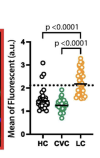
We observed that purified total IgG from a subset of LC participants showed increased reactivity against human locus coeruleus (Fig.1B), human thalamus (Fig. 1C), mouse sciatic nerve (Fig. 1D), and mouse meninges (Fig. 1E). Few participants also displayed positivity against human dorsolateral prefrontal cortex (Supp. Fig.1B).

Human Locus coeruleus Fig 1 B.......Human thalamus Fig 1C..... Mouse sciatic nerve Fig 1D ,,,,,,,, Mouse meninges Fig 1E
Screenshot 2026-05-29 at 9.02.49 AM.png ........ Screenshot 2026-05-29 at 9.04.06 AM.png,,,,,,,,,,,,,,,,,Screenshot 2026-05-29 at 9.05.48 AM.png..........Screenshot 2026-05-29 at 9.06.49 AM.png

Sorry for the less than wonderful formatting, Those results aren't all that compelling, except for the mouse meninges. But, I wonder how many tissues did they test - there may have been a lot given there are human and mice tissues? And what did the chart for human meninges look like? Are there charts for tissues where the healthy controls and convalescent controls have more fluorescence that have not been presented in the paper? (I haven't checked the methods section.)

In general, the staining presented a diffuse pattern, with few cells demonstrating distinct labeling (Supp. Fig. 2 and Supp. Fig. 1C). On average 93.94% of healthy and 85.71% of convalescent controls tested positive for at least one neurological tissue. At the same time, long COVID patients exhibited reactivity to an average of 3.6 human neurologic tissues per individual, compared to healthy control (2.4 tissues) and convalescent control (2.5 tissues) (Supp. Fig. 1A).
Note that they don't tell us what percentage of LC people tested positive for at least one neurological tissue. Almost all of the controls tested positive for reactivity to one neurological tissue. Those differences in the number of tissues where reactivity was found are not compelling.

This is about periperhal tissues:
We observed that on average 52.17% of the Long COVID individuals were positive to at least one human peripheral tissue, with a mean of 3.41 tissues per individual (Supp. Fig. 3A). Individuals with Long COVID showed diverse and increased positivity to several human peripheral tissues, including the adrenal gland, heart muscle, parathyroid, and thyroid tissues (Supp. Fig. 3B). A representative staining pattern for specific tissues are depicted in Supp. Fig. 2 and Supp. Fig 4. The number of positive tissues per individual with Long COVID is described in Supp. Fig. 3A.
Note there that they don't tell us what percentage of the controls were positive to at least one human peripheral tissue. I assume they would if there were compelling differences. It's coming across as rather underwhelming.

Thus, Long COVID patients have a diverse and increased number of autoantibodies against human peripheral and CNS tissues. .....ROC analysis demonstrated good discriminatory capacity between LC and controls for locus coeruleus and mouse meninges, supporting the robustness of the IF-based findings (Fig.1B and Fig. 1E). ROC analysis for all tissues is shown in Supp. Fig. 1 and Supp. Fig. 3.
To account for age and sex as a confounder in our analysis, we performed a linear regression analysis adjusting for age and sex (Supp. Fig. 5I). Notably, only mouse meninges remained significant despite several tissues showing an overall increase in autoreactivity in the LC group. We stained meninges from both female and male mice to compare sex-related differences in staining patterns in a sample classified as positive but observed no differences (Supp. Fig. 1D).
It's good that they did that adjustment for age and sex confounding. But, for only one (mouse) tissue to remain significant when they tested so many tissues - well, it doesn't seem very convincing.

I haven't read the whole paper, I haven't chased down every figure in the supplementary material and all the information in the Methods section. Perhaps the AABs are more harmful in LC than in the controls? I'd really love for AABs to be the problem causing our illnesses. But, this part of the paper doesn't look strong. The work doesn't seem to have been presented in its totality.

And, if AABs were the problem, then I think we'd know that IVIG cures us? I don't think we know that.
 
Back
Top Bottom