Wyva
Senior Member (Voting Rights)
Donna M. Mancini, Danielle L. Brunjes, Dane Cook, Tiffany Soto, Michelle Blate, Patrick Quan, Tadahiro Yamazaki, Anna Norweg, Benjamin H. Natelson
Abstract
Introduction: Patients with myalgic encephalomyelitis/chronic fatigue syndrome (ME/CFS) experience symptoms of fatigue, dyspnea, mental fog, and worsening fatigue after physical or mental efforts. Some of these patients have been found to hyperventilate. In long COVID patients, many of whom also have ME/CFS, dysfunctional breathing (DB) has been described. Whether patients with ME/CFS, independent of COVID-19, experience dysfunctional breathing is unknown, as well as how it may relate to hyperventilation.
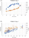
Methods: We performed serial 2-day cardiopulmonary exercise testing (CPET) in 57 patients with ME/CFS and 25 age- and activity-matched control participants. Peak oxygen consumption (VO2), ventilatory efficiency slope (VE/VCO2), O2 saturation, end-tidal CO2 (PetCO2), heart rate, and mean arterial blood pressure were measured in all patients during upright incremental bicycle exercise. Ventilatory patterns were reviewed using minute ventilation (VE) versus time, respiratory rate, and tidal volume versus minute ventilation graphs. Chronic hyperventilation (HV) was defined as a PETCO2 of <34 mm Hg that persisted during low-intensity exercise. Dysfunctional breathing was characterized by a 15% increase in oscillations in minute ventilation during at least 60% of the exercise duration or by a scatterplot pattern of respiratory rate and tidal volume plotted versus minute ventilation.
Results: The patients with ME/CFS had an average age of 38.6 ± 9.6 years, and a mean body mass index (BMI) of 24.1 ± 3.4, which was comparable to the sedentary controls. All participants performed maximal exercise, achieving a respiratory exchange ratio (RER) of >1.05. For the patients with ME/CFS, peak VO2 averaged 22.3 ± 5.3 mL/kg/min, which was 79 ± 20% of predicted and comparable to that observed in the sedentary controls (23.4 ± 4.6 mL/kg/min; 81 ± 12%; p = NS). A total of 24 patients with ME/CFS (42.1%) met the criteria for dysfunctional breathing compared to four sedentary controls (16%) (p < 0.02). In total, 18 patients with ME/CFS (32%) had hyperventilation compared to one sedentary control participant (4%) (p < 0.01), and nine patients with ME/CFS had both hyperventilation and dysfunctional breathing, whereas no sedentary participant exhibited both. The patients with ME/CFS and hyperventilation had significantly higher VE/VCO2 ratios (HV+: 34.7 ± 7.2; HV−: 28.1 ± 3.8; p < 0.001). A total of 15 of 18 patients with hyperventilation (83%) had either elevated VE /VCO2 ratios (n = 15) or dysfunctional breathing (n = 9) compared to 44% (n = 17) of the 40 non-hyperventilators (p < 0.01).
Conclusion: Dysfunctional breathing and hyperventilation are common in patients with ME/CFS and could present a new therapeutic target for these patients.
Open access: https://www.frontiersin.org/journals/medicine/articles/10.3389/fmed.2025.1669036/full


