Cellular immune function in myalgic encephalomyelitis/chronic fatigue syndrome (ME/CFS) (2019) Cliff, Nacul et al.

Cheshire

Senior Member (Voting Rights)
Myalgic encephalomyelitis/chronic fatigue syndrome (ME/CFS) is a debilitating condition with unknown aetiology, unclear pathophysiology and with no diagnostic test or biomarker available. Many patients report their ME/CFS began after an acute infection, and subsequent increased frequency of infections, such as colds or influenza, is common. These factors imply an altered immunological status exists in ME/CFS, in at least a proportion of patients, yet previous studies of peripheral immunity have been discrepant and inconclusive.

The UK ME/CFS Biobank, which has collected blood samples from nearly 300 clinically-confirmed ME/CFS patients, enables large-scale studies of immunological function in phenotypically well-characterised participants. In this study, herpes virus serological status and T cell, B cell, NK cell and monocyte populations were investigated in 251 ME/CFS patients, including 54 who were severely affected, and compared with those from 107 healthy participants and with 46 patients with Multiple Sclerosis.

There were no differences in seroprevalence for six human herpes viruses between ME/CFS and healthy controls, although seroprevalence for the Epstein-Barr virus was higher in multiple sclerosis patients. Contrary to previous reports, no significant differences were observed in NK cell numbers, subtype proportions or in vitro responsiveness between ME/CFS patients and healthy control participants.

In contrast, the T cell compartment was altered in ME/CFS, with reduced proportions of effector memory CD8+ T cells and of intermediately differentiated CD8+ T cells in ME/CFS. Conversely, there was a significantly increased proportion of mucosal associated invariant T cells (MAIT) cells, especially in severely affected ME/CFS patients.

These abnormalities demonstrate that an altered immunological state does exist in ME/CFS, particularly in severely affected people. This may simply reflect ongoing or recent infection, or may indicate future increased susceptibility to infection. Longitudinal studies of ME/CFS patients are needed to help to determine cause and effect and thus any potential benefits of immuno-modulatory treatments for ME/CFS.

https://www.frontiersin.org/articles/10.3389/fimmu.2019.00796/abstract

Full, open access, text here, https://www.frontiersin.org/articles/10.3389/fimmu.2019.00796/full

ETA: Link to full text.
 
Last edited by a moderator:
Very interesting study and i look forward learning more. Point of interest, the mention on MAIT Cells :


In contrast, the T cell compartment was altered in ME/CFS, with reduced proportions of effector memory CD8+ T cells and of intermediately differentiated CD8+ T cells in ME/CFS. Conversely, there was a significantly increased proportion of mucosal associated invariant T cells (MAIT) cells, especially in severely affected ME/CFS patients.

A paper i found named "Interactions between bile salts, gut microbiota, and hepatic innate immunity" mentions :


Liver lymphocytes consist of B cells, conventional T cells (viz. T-helper cells, regulatory T cells, and cytotoxic T cells), and unconventional T cells like natural killer (NKT) T cells and mucosal-associated invariant T (MAIT) cells.80

The latter are an emerging type of unconventional, innate-like T cells that are highly abundant in the human liver where they comprise up to 50% of total T cells.81 MAIT cells may play a role in the immunological homeostasis of the liver. They are also abundant in the gut lamina propria 82 and represent 1-10% of CD3+ T cells in the peripheral blood

I wonder if this has anything to do with what was found. Let's not also forget about the -recent- paper by Professor Tate in New Zealand (cc @Hutan ) that suggests inflammation of the liver given the pathways found:


latest-png.5574
 
From the abstract:
”Many patients report their ME/CFS began after an acute infection, and subsequent increased frequency of infections, such as colds or influenza, is common”

I do not think this is quite accurate from my understanding of how ME evolves. Yes, patients get initial infection, but what follows is a period of time, often years when patients do not catch a cold or get sick with common viral infections at all. The immune system seems to be on high alert all the time.

Then after that period of over active immune system, in my case 10 years it is as if the immune system gets depleted and patients are prone to such infections such as colds and flus.

Dr Lipkin and team (including CFI) examined that concept by assessing patients in early stages of disease vs late stages.
 
I think it's different by patient whether we seem more susceptible to incidental infections, less susceptible, or just seem to get things but not get all the associated symptoms. It does seem to change over time for some.

I caught everything in my early years of being ill, especially if I was not rigorous about hand hygiene. This changed not to not getting things, but to not having all the symptoms when I do. (for example, I rarely run a fever even when having an infection that would normally produce a fever--however doctors rarely listen to me when I tell them they can't use absence of fever as a decision aid)
 
Lay summary: Increased MAIT cell frequency in ME/CFS

Immune system dysfunction has long been suspected in ME/CFS, with many patients reporting an infection preceding their illness, as well as increased frequency of infections following disease onset. Yet, the results of immune studies in ME/CFS have been conflicting and inconclusive.

In our recent publication, we looked at several aspects of immune function in the blood cells of 251 people with ME/CFS, including 54 who were severely affected, and compared these with 107 healthy controls and 46 people with MS (Multiple Sclerosis).

Firstly, we measured the presence of antibodies against six different Herpes viruses in the blood, including Epstein-Barr virus, and found no difference between ME/CFS patients and controls. Next, we looked at different immune cells, including T cells and NK cells. Contrary to previous findings, no differences were found in NK cells between ME/CFS patients and controls. We also found no differences in the overall number of T cells, although differences were found in the proportions of different types of T cells in ME/CFS patients compared to controls; the functional role of these differences is yet to be determined.
https://cureme.lshtm.ac.uk/laboratory-results-published/
 
Sample Collection and Processing, Including PBMC Freezing
...
Samples were delivered to the University College London / Royal Free Hospital (UCL/RFH) BioBank laboratories for processing and storage within 6 h of collection: the average time from collection to processing was 2.5 h across all four study groups.

Serum, plasma, and peripheral blood mononuclear cells (PBMC) were processed and aliquoted in volumes of 200 μl to 2 ml. PBMCs were aliquoted at 5 × 10⁶ cells/ml. Aliquots were stored in 1 or 2 ml cryotubes, in vapour phase liquid nitrogen at −180°C, and shipped to LSHTM for laboratory analysis on dry ice.

This could be why have a different finding with NKCC. Some of the other labs are not freezing their samples.

Freezing is a potential source of differences in findings.
https://www.ncbi.nlm.nih.gov/pmc/articles/PMC3400260/
 
Within the leucocyte gate, monocytes were identified by CD14 positivity and CD3 negativity
Therefore I think non-classical monocytes maybe were not investigated in this study, certainly not individually.
Human monocytes are divided in three major classes; classical (CD14+CD16−), non-classical (CD14dimCD16+), and intermediate (CD14+CD16+).

This study has been cited as evidence that the increase in non-classical monocytes that has been found in two studies of Long Covid patients has not been found in ME/CFS. I don't think this is correct, as the non-classical monocytes subset wasn't specifically looked at.
Persistence of SARS CoV-2 S1 Protein in CD16+ Monocytes in Post-Acute Sequelae of COVID-19 (PASC) ..., 2021, Patterson et al (in prep)
 
Last edited:
Adding link to post containing video of Dr Jaqueline Cliff's presentation posted March 2025 that builds on the work on this paper. In the video Dr Cliff presents new data that just like this one shows T cell behaviour differs between those with Severe vs Mild/Moderate ME/CFS
Post : https://www.s4me.info/threads/updates-from-the-uk-me-cfs-biobank-cureme-team.2401/page-6#post-593498
Video : https://cureme.lshtm.ac.uk/index.php/2025/03/10/first-cure-me-webinar/

I think this 2019 paper may be worth revisiting in combination with other papers comparing severe vs mild/moderate ME to build a list of all the differences that have been found between the two sets.
Tagging @Murph as you were keen to revisit data in existing papers to see if anything new could be learned.
Tagging @mariovitali for same reason.
List of Cure ME Biobank papers can be found on the Cure ME website and the NIH publication list for this team. Many of these papers separate data for the two groups.
 
Thinking about this paper and the presentation @wigglethemouse highlighted in the context of the expanded T cell populations and if how expanded they are correlates with severity. Haven’t got much further but would be interested in others thoughts.
 
Thinking about this paper and the presentation @wigglethemouse highlighted
For reference, Jackie Cliff recently presented some findings on the latest work at a CureME webinar earlier this year (Link to post with video).

I wondered what could be the reason severe ME patients do not show a correlation between anti-HHV6B antibodies and cytotoxic Tcell activity but mild/moderate patients do. (Link to post).

Haven’t got much further but would be interested in others thoughts.
I think it would be good to revisit this paper along with the data in the webinar from Jackie Cliff to see if what we can (re-)learn. The paper of this thread did the work on frozen cells, and the Selin lab works with fresh cells with different sorting methods, which led to some differences on double positive T-cells for example.
 
Small changes in ME/CFS T-cells compared to other groups. I've no idea what these cell types are (EM & TEMRA cells). [No figure included here because it is not legible].
However, modest differences were observed in several differentiation populations in the CD8+ T cell compartment (Figure 5B) with a small increase in the proportion of EM cells (CCR7-CD45RA-CD28-CD57-) and a reduction in the proportion of terminally differentiated cytotoxic effector TEMRA cells (CCR7-CD45RA+CD28–CD57+) in people living with ME/CFS.

There were also small changes in other minor CD8+ T cell populations, with small reductions in proportions of CCR7-CD45RA+CD28+CD57+, CCR7+CD45RA-CD28+CD57-, and CCR7-CD45RA-CD28+CD57- cells in people with ME/CFS and an increased proportion of CCR7+CD45RA+CD28-CD57- cells.

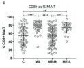
It seems MAIT cells that express CD8 are more common in severe ME/CFS, similar to that seen in MS, but different from healthy controls.

1752000079078.png
Of greater potential relevance, however, using TCR Vα7.2 and CD161 to define circulating populations of mucosal associated invariant T (MAIT) cells, we observed highly significant differences between the groups in the proportion of T cells that are MAIT cells and the proportion of MAIT cells that express CD8 (unadjusted ANOVA, P < 0.001 in both cases) (Figure 6).

After adjusting for confounding, the multiple linear analysis revealed that this was due to increased proportions of MAIT cells (P < 0.001), and particularly of CD8+ MAIT cells, in people with severe ME/CFS compared to healthy controls (P < 10−5).

Importantly however, although the overall proportions of MAIT cells were not higher among people with MS than among healthy controls, their MAIT cells were also heavily skewed to the CD8+ subset, indicating that this is not a diagnostic feature of severe ME/CFS cases.

Potentially more importantly, the proportion of MAIT cells which were CD8+ (Figure 6G) had a greater capacity to discriminate severe ME/CFS from health, with an AUC of 0.756.
 
For those interested in IFNγ there was a difference in CMV seropositive individuals, but it was across all groups. Does this mean in the JE hypothesis we might expect enhanced effect in those with CMV seropositivity despite no relation to severity or could this be some negative data against the hypothesis?
We therefore compared lymphocyte phenotype and function among those subjects who were either seropositive or not for CMV. Across all the study groups, participants who were CMV-seropositive had significantly lower proportions of CD4+ and higher proportions of CD8+ T cells amongst their PBMC (Supplementary Figure S5).

Following stimulation with PMA and ionomycin, higher proportions of both CD4+ and CD8+ T cells made IFNγ, and more cells were IL-2 and IFNγ double positive, amongst CMV seropositive individuals.

Amongst CMV-seropositive individuals, there was a decreased in vitro response to IL-12 and IL-18 stimulation, with significantly reduced CD25, CD107, and IFNγ expression (Supplementary Figure S5G).
 
Back
Top Bottom