Folliculostellate (FS) Cells

Here’s a comparison of some mentioned marker genes expressed on astrocytes and FS cells from data from the human protein atlas. Lots of similarities, and now HLA, but the CD74 may be an interesting one considering the IFN gamma involvement?

Code:
================================================================================
HPA SINGLE-CELL GENE EXPRESSION SUMMARY (Expression Metric: NCPM)
================================================================================
gene name                   Category  cell type   ncpm
    S100B   FS / Epithelial Identity astrocytes 256.40
     SOX2   FS / Epithelial Identity astrocytes   2.10
    PTPRC Leukocyte / Myeloid Marker astrocytes   3.10
     CD14 Leukocyte / Myeloid Marker astrocytes   0.90
    HLA-A MHC Class I (Constitutive) astrocytes  44.60
    HLA-C MHC Class I (Constitutive) astrocytes  33.40
    HLA-E MHC Class I (Constitutive) astrocytes  21.70
    HLA-B MHC Class I (Constitutive) astrocytes  12.70
    HLA-F MHC Class I (Constitutive) astrocytes   5.80
 HLA-DPB1   MHC Class II (Inducible) astrocytes   3.50
  HLA-DRA   MHC Class II (Inducible) astrocytes   1.70
 HLA-DRB1   MHC Class II (Inducible) astrocytes   1.60
 HLA-DPA1   MHC Class II (Inducible) astrocytes   1.20
 HLA-DQB1   MHC Class II (Inducible) astrocytes   0.20
 HLA-DQA1   MHC Class II (Inducible) astrocytes   0.20
     CD74     MHC Class II Accessory astrocytes   4.30
    CIITA      Master Transactivator astrocytes   3.20


Code:
================================================================================
HPA SINGLE-CELL GENE EXPRESSION SUMMARY (Expression Metric: NCPM)
================================================================================
gene name                   Category       cell type  ncpm
    S100B   FS / Epithelial Identity pituicytes/fscs 64.50
     SOX2   FS / Epithelial Identity pituicytes/fscs  0.50
     CD14 Leukocyte / Myeloid Marker pituicytes/fscs  1.30
    PTPRC Leukocyte / Myeloid Marker pituicytes/fscs  0.80
    HLA-A MHC Class I (Constitutive) pituicytes/fscs 59.60
    HLA-C MHC Class I (Constitutive) pituicytes/fscs 11.20
    HLA-F MHC Class I (Constitutive) pituicytes/fscs  4.90
    HLA-B MHC Class I (Constitutive) pituicytes/fscs  3.80
    HLA-E MHC Class I (Constitutive) pituicytes/fscs  1.40
 HLA-DRB1   MHC Class II (Inducible) pituicytes/fscs  6.30
 HLA-DQB1   MHC Class II (Inducible) pituicytes/fscs  5.10
 HLA-DPB1   MHC Class II (Inducible) pituicytes/fscs  4.00
 HLA-DQA1   MHC Class II (Inducible) pituicytes/fscs  0.70
  HLA-DRA   MHC Class II (Inducible) pituicytes/fscs  0.60
 HLA-DPA1   MHC Class II (Inducible) pituicytes/fscs  0.40
     CD74     MHC Class II Accessory pituicytes/fscs 40.40
    CIITA      Master Transactivator pituicytes/fscs  4.60
These cells, their function w.r.t. cognition and the links to IFNy, innate immunity etc are really interesting. But based on these marker gene associations, would we not have seen a clear HLA signal in DecodeME if they were involved?
 
Last edited:
These cells, their function w.r.t. cognition and the links to IFNy, innate immunity etc are really interesting. But based on these marker gene associations, would we not have seen a clear HLA signal in DecodeME if they were involved?

I don’t think we’ll see hits for every gene for every protein or receptor for every cell involved in whatever process we uncover. It’s also worth saying these FS cells aren’t showing high expression, just some, it’s that and the patterns and differences which I’m assuming probably account for some of the behaviours seen. Would be interested in what others think though.

There may of course be no relevance here ofc. I’m just going through some mechanisms, learning what I can there’s a few ways I can see them being involved but it doesn’t necessarily mean they are! It’s interesting though and I hope these posts are bringing some others interested along on the journey.
 
Hopefully this next bit will help with the wider picture and help explain a bit we’ve covered how these calcium waves propagate through the network but what triggers those calcium waves and what could alter how of when they are?

First Il-6. This confused me a lot as there are clear claims that FS cells produce IL-6. But looking at the papers this is not in humans, rather/mouse models.

Production of Interleukin-6 by Folliculo-Stellate Cells of the Anterior Pituitary Gland in a Histiotypic Cell Aggregate Culture System, 2008, Vankelecom et al

Reaggregate cell cultures of mouse or rat anterior pituitary were found to produce interleukin-6 (IL-6)

Studies on aggregates prepared from differentially enriched pituitary cell populations revealed the presence of folliculo-stellate (FS) cells to be essential for IL-6 production.

Aggregates that contained only hormone-secreting, but no FS cells, failed to produce IL-6. Furthermore, the yield of IL-6 increased with increasing proportions of FS cells present in the aggregates.

Web | DOI | PDF | Neuroendocrinology

Rather than quoting lots of papers I’ll try to cover what I think the situation is how I reconciled it in my head and others can judge.

Accessing live human pituitary tissue presents understandable challenges. There are studies from autopsies/tumours but there are technical issues around staining and tissue degradation and tumours behave differently with different sources of IL-6. And things just moved on from tissue sampling to RNA sequencing.

Human protein atlas data for Il6 for the pituitary shows the following

Average nTPM: 39.0

Samples: 313
Max nTPM: 736.5

Min nTPM: 0.0

Std nTPM: 97.8

Median nTPM: 4.1

And perhaps more convincingly single cell RNA expression shows this is almost entirely in Pituicytes/FSCs rather than lactotrophs, somatotrophs, macrophages, etc.

Of note the data doesn’t distinguish between anterior and posterior, so we cannot be sure it’s pituicytes or FS cells or both and there is quite a range of values in the samples. But it seems consistent with the claims in other papers and the findings in rodents that FS cells produce IL-6.

 
Last edited:
Hopefully this next bit will help with the wider picture and help explain a bit we’ve covered how these calcium waves propagate through the network but what triggers those calcium waves and what could alter how of when they are?

First Il-6. This confused me a lot as there are clear claims that FS cells produce IL-6. But looking at the papers this is not in humans, rather/mouse models.

Production of Interleukin-6 by Folliculo-Stellate Cells of the Anterior Pituitary Gland in a Histiotypic Cell Aggregate Culture System, 2008, Vankelecom et al

Reaggregate cell cultures of mouse or rat anterior pituitary were found to produce interleukin-6 (IL-6)

Studies on aggregates prepared from differentially enriched pituitary cell populations revealed the presence of folliculo-stellate (FS) cells to be essential for IL-6 production.

Aggregates that contained only hormone-secreting, but no FS cells, failed to produce IL-6. Furthermore, the yield of IL-6 increased with increasing proportions of FS cells present in the aggregates.

Web | DOI | PDF | Neuroendocrinology

Rather than quoting lots of papers I’ll try to cover what I think the situation is how I reconciled it in my head and others can judge.

Accessing live human pituitary tissue presents understandable challenges. There are studies from autopsies/tumours but there are technical issues around staining and tissue degradation and tumours behave differently with different sources of IL-6. And things just moved on from tissue sampling to RNA sequencing.

Human protein atlas data for Il6 for the pituitary shows the following

Average nTPM: 39.0

Samples: 313
Max nTPM: 736.5

Min nTPM: 0.0

Std nTPM: 97.8

Median nTPM: 4.1

And perhaps more convincingly single cell RNA expression shows this is almost entirely in Pituicytes/FSCs rather than lactotrophs, somatotrophs, macrophages, etc.

Of note the data doesn’t distinguish between anterior and posterior, so we cannot be sure it’s pituicytes or FS cells or both and there is quite a range of values in the samples. But it seems consistent with the claims in other papers and the findings in rodents that FS cells produce IL-6.
IL-6 has come up a few times in the research no? I think in long covid in particular
 
IL-6 has come up a few times in the research no? I think in long covid in particular
I’m not sure! You have a far better memory of these things. And the more I look at some of this the more possibilities I see but also the less sure of some other things I become!

Some people/papers talk about the pituitary being at the crossroads of the immune, hormone and nervous systems so I wondered if there was something here. Maybe it could help explain or tie up with the prolactin findings but I’m not sure tbh.
 
m not sure! You have a far better memory of these things. And the more I look at some of this the more possibilities I see but also the less sure of some other things I become!
It's too short a term to search on S4ME but I think raised IL-6 was the basis for the tocilizumab trial in the UK and has been seen up in some long covid studies.

[Edit: Tocilizumab study:

]


Some people/papers talk about the pituitary being at the crossroads of the immune, hormone and nervous systems so I wondered if there was something here. Maybe it could help explain or tie up with the prolactin findings but I’m not sure tbh
It does seem like all this could tie together really well. No guarantee that it means anything ofc but it would answer a lot of questions about the quirks of ME/CFS symptoms if the answer (or part of it) was to do with FS cells and the pituitary.
 
Last edited:
It does and it would. You’re not the only one who sees things everywhere :) For instance there is this paper which would maybe tie in with some innate immune angles (LPS, IL-6, TLR4, tick, tick, tick). But human protein atlas data shows it’s mainly macrophages which express CD14 in the pituitary not FS cells and there’s no sign of TLR4. So maybe it’s just not transferable to humans?
Lipopolysaccharide Directly Stimulates the Intrapituitary Interleukin-6 Production by Folliculostellate Cells via Specific Receptors and the p38α Mitogen-Activated Protein Kinase/Nuclear Factor-κB Pathway**Supported by a grant from the DFG: Sta 285/7–3., 2000, Lohrer et al

Lohrer, P.; Gloddek, J.; Nagashima, A. Carbia; Korali, Z.; Hopfner, U.; Pereda, M. Paez; Arzt, E.; Stalla, G. K.; Renner, U.


Bacterial lipopolysaccharide (LPS) activates the immune system and induces increases in peripheral cytokines, which, in turn, affect the endocrine system. In particular, LPS-induced cytokines stimulate the hypothalamic-pituitary-adrenal axis to increase levels of antiinflammatory-acting glucocorticoids.

In the present work, we show that LPS directly stimulates interleukin (IL)-6 release by mouse pituitary folliculostellate (FS) TtT/GF tumor cells and FS cells of mouse pituitary cell cultures.

Both TtT/GF cells and mouse pituitaries expressed CD14, which binds the LPS/LBP complex, and Toll-like receptor type 4, which induces LPS signals. LPS increased phospoinositol turnover in TtT/GF cells and induced phosphorylation of p38alpha mitogen-activated protein kinase and the inhibitor (IkappaB) of nuclear factor-kappa B. Nuclear factor-kappa B was activated by LPS in TtT/GF cells.

Functional studies demonstrated that My4 (an antibody blocking the interaction between LPS/LBP and CD14), SB203580, (a specific inhibitor of p38alpha mitogen-activated protein kinase phosphorylation), dexamethasone, and the messenger RNA translation inhibitor cycloheximide all inhibited LPS-induced IL-6 production by TtT/GF cells and mouse pituitary FS cells. LPS-induced intrapituitary IL-6 may modulate the function of anterior pituitary cells during bacterial infection/inflammation.

Web | DOI | Endocrinology
 
Last edited:
Thanks for the study info @V.R.T. Looks like they’re maybe going the autoimmunity line of thinking? What I was wondering here was more like a localised messenger role. It’s not that the FS cells produce loads of cytokines that would cause systemic issues. More they seem to stimulate release of hormones. Another old/rodent paper

Interleukin 6 possibly induced by interleukin 1β in the pituitary gland stimulates the release of gonadotropins and prolactin, 1990, Yamaguchi et al

The abilities of recombinant human interleukin 1 (IL-1) and IL-6 to induce release of FSH, LH and PRL from rat pituitary cells in vitro were examined. IL-1 and IL-6 induced significant releases of FSH, LH and PRL within 3 h. The extents of release of these compounds induced by IL-1 and IL-6 were similar to those induced by GnRH and TRH.

Rat anterior pituitary cells released IL-6 spontaneously, and its release was enhanced by IL-1β. This effect of IL-1β was inhibited significantly by a rabbit anti-IL-1β antiserum.

These findings suggest that IL-1 induced the release of IL-6 from rat pituitary, and that the released IL-6 stimulated the secretions of FSH, LH and PRL.

Web | DOI | Acta Endocrinologica
 
Last edited:
This was also interesting. I’m finding most mechanistic studies are not in humans. Because of the challenges getting healthy live human pituitary tissue most experiments appear to be in rodent models. To try to deal with some of the differences this study inserts human gene fragments into rodent cell lines.

Human Prolactin Gene Promoter Regulation by Estrogen: Convergence with Tumor Necrosis Factor-α Signaling, 2007, Adamson et al

Estrogens have been implicated in the regulation of prolactin gene expression in man, although previous studies have not defined the molecular mechanism whereby estradiol activates the human prolactin gene promoter (hPrl). We found that estradiol induced a reproducible 1.8-fold activation of the hPrl gene promoter, using pituitary GH3 cells stably transfected with a 5000-bp hPrl promoter fragment linked to luciferase reporter gene. This activation was blocked by treatment with estrogen receptor (ER) antagonists 4-hydroxytamoxifen and ICI-182,780.

Promoter deletion and mutagenesis experiments identified a functional estrogen response element (ERE) sequence 1189 bp upstream of the transcription start site that was responsible for estrogen-mediated promoter activation. This site differed from the consensus ERE sequence by two base pairs, one in each half-site. This ERE was identified to be functional through binding ERα in EMSAs. Chromatin immunoprecipitation assays confirmed ERα binding to this sequence in vivo in the absence of ligand, with increased recruitment when cells were cultured in the presence of estradiol.

When cells were treated with both estradiol and TNFα, we observed synergistic activation of the hPrl promoter, which was mediated by the −1189-bp ERE. Mutagenesis of this ERE abolished the promoter-activating effect not only of estradiol but also of TNFα.

These data suggest a novel, promoter-specific signaling interaction between estrogen and TNFα signaling, which is likely to be important for prolactin regulation in vivo.

Web | DOI | PMC | PDF | Endocrinology

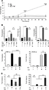
So I think in this paper they built upon some other papers of the time and found (1) estradiol increased activation of the prolactin gene promoter (2) identified a section of the dna that appeared to be responsible for this (3) found that estradiol and TNFα worked together at this section and (4) that disrupting this section stopped the effects.

Here’s the figure showing the individual and combined effects over time

IMG_4180.webp
 
Last edited:
So again moving to protein atlas data, what do we see in human tissue samples (noting this a measure of normalised transcript expression, not a measure of what’s always happening in a cell, but it can give us an idea of what a cell is likely capable of).

Data doesn’t support any ideas that TNF-alpha is produced locally by FS cells or lactotrophs, so any signal is coming from somewhere else. But the values for NFKB1 (along with RIPK1 and ADAM10) seems to support the findings of the paper that shows TNF-alpha can enhance production of prolactin in lactotrophs, either alone or in concert with estrogen. Because those are used by cell machinery in TNF-alpha related signalling and processing (although not exclusively, they are also used in other things). So human lactotrophs seem to have the machinery to respond to TNF-alpha signals.

Perhaps interestingly, in the data lactotrophs don’t show much in the way of TNF-Alpha receptor (TNFRSF1A or TNFRSF1B) expression though. And the precursor paper to the above (Tumor Necrosis Factor-α Activates the Human Prolactin Gene Promoter via Nuclear Factor-κB Signaling, 2006, Friedrichsen et al) particular focused on this as a route and receptor activity would be expected for direct interaction.

This is where FS cells may come in as they posses the lot. They appear to have the machinery to process and signal but also the receptors. This raises the possibility that FS cells could act as intermediaries in some way.

Code:
HPA SINGLE-CELL GENE EXPRESSION SUMMARY (Expression Metric: NCPM)

=======================================================================

=========

gene name                  Category       cell type   ncpm

    S100B Identity / Lineage Marker pituicytes/fscs  64.50

     CD14 Identity / Lineage Marker pituicytes/fscs   1.30

    PTPRC Identity / Lineage Marker pituicytes/fscs   0.80

     SOX2 Identity / Lineage Marker pituicytes/fscs   0.50

   NFKBIA         Negative Feedback pituicytes/fscs  41.40

  TNFAIP3         Negative Feedback pituicytes/fscs   3.10

    NFKB1       Receptor Signalling pituicytes/fscs 131.40

    RIPK1       Receptor Signalling pituicytes/fscs  37.70

     RELA       Receptor Signalling pituicytes/fscs  18.40

    TRADD       Receptor Signalling pituicytes/fscs   6.80

    TRAF2       Receptor Signalling pituicytes/fscs   4.70

      LTA   TNF Ligand (Production) pituicytes/fscs   0.00

      LTB   TNF Ligand (Production) pituicytes/fscs   0.00

      TNF   TNF Ligand (Production) pituicytes/fscs   0.00

   ADAM17 TNF Processing / Shedding pituicytes/fscs 223.20

   ADAM10 TNF Processing / Shedding pituicytes/fscs  89.60

TNFRSF1A              TNF Receptor pituicytes/fscs  93.40

TNFRSF1B              TNF Receptor pituicytes/fscs   4.20


Code:
HPA SINGLE-CELL GENE EXPRESSION SUMMARY (Expression Metric: NCPM)

================================================================================

gene name                  Category   cell type   ncpm

    S100B Identity / Lineage Marker lactotrophs   2.10

     CD14 Identity / Lineage Marker lactotrophs   1.50

    PTPRC Identity / Lineage Marker lactotrophs   1.10

     SOX2 Identity / Lineage Marker lactotrophs   0.00

   NFKBIA         Negative Feedback lactotrophs  15.60

  TNFAIP3         Negative Feedback lactotrophs   0.50

    NFKB1       Receptor Signalling lactotrophs 113.10

    RIPK1       Receptor Signalling lactotrophs  46.00

    TRAF2       Receptor Signalling lactotrophs  15.50

     RELA       Receptor Signalling lactotrophs   7.20

    TRADD       Receptor Signalling lactotrophs   5.50

      LTA   TNF Ligand (Production) lactotrophs   0.00

      LTB   TNF Ligand (Production) lactotrophs   0.00

      TNF   TNF Ligand (Production) lactotrophs   0.00

   ADAM10 TNF Processing / Shedding lactotrophs 154.60

   ADAM17 TNF Processing / Shedding lactotrophs  83.10

 TNFRSF1A              TNF Receptor lactotrophs   8.20

 TNFRSF1B              TNF Receptor lactotrophs   0.90
 
Last edited:
And then there’s this, something that ties together calcium waves and the coordinated pulses of the earlier reviews with the cytokines above? More to think about although for now my brain is fried. But the result could be some sort of negative feedback loop or maybe just a noisy and less coordinated network when? Again this is in rodents, but some interesting possibilities!

Tumor Necrosis Factor-α-Induced Anterior Pituitary Folliculostellate TtT/GF Cell Uncoupling Is Mediated by Connexin 43 Dephosphorylation, 2007, Meilleur et al

The anterior pituitary folliculostellate (FS) cells are key elements of the paracrine control of the pituitary function. These cells are the source and the target of growth factors and cytokines, and are connected to other pituitary cells via Cx43-mediated gap junctions.

Here, we show that acute treatment of the FS TtT/GF cell line with TNF-α caused a transient cell uncoupling that was accompanied by the dephosphorylation of Cx43 in Ser368. These TNF-α-evoked effects were dependent on protein phosphatase 2A (PP2A) and protein kinase C (PKC) activities.

Moreover, this interaction was weakened by TNF-α.

The temporary closure of gap junctions during acute TNF-α challenge may constitute a protective mechanism to limit or confine the spread of inflammatory signals among the FS cells.

https://academic.oup.com/endo/article/148/12/5913/2501831']Web | DOI | PDF | Endocrinology

Human protein atlas data shows GJA1 (which encodes connexin 43/Cx43) is present in FSCs and vascular endothelial cells in the pituitary.
 
One aspect that perhaps needs highlighting from the earlier paper on gap junctions and calcium waves and also supported by others is that the connections are not just between FS cells but also between FS cells and hormone producing cells, particularly lactotrophs (PRL) and somatotrophs (GH). As an aside, I understand lactotrophs and somatotrophs also share a developmental lineage.

Cell-to-cell communication in the anterior pituitary: evidence for gap junction-mediated exchanges between endocrine cells and folliculostellate cells., 1996, Morand et al

The ability of rat anterior pituitary cells to communicate through gap junctions (GJ) was studied using a fluorescent molecule, Lucifer Yellow (LY), which freely passes through GJ channels.
The identification of communicating cells was performed by immunofluorescence labeling of specific hormones in endocrine cells and of S100 protein in folliculostellate (FS) cells. Rat anterior pituitary cells in their physiological organization, i.e. in the intact tissue, exhibited a high level of coupling through GJ.
The communicating cells were primarily PRL cells, GH cells, and FS cells. Only a few LH, TSH, and ACTH cells were labeled with LY.
By immunolabeling of specific hormones and/or S100 protein, we found a GJ coupling between FS cells, and between FS cells and endocrine cells, including PRL cells. The communication between FS cells was by far the most frequent.
In conclusion, we demonstrate the presence of functional GJ between anterior pituitary cells of the same type and between anterior pituitary cells having distinct differentiated functions.

Web | DOI | Endocrinology

Putting this together with earlier papers we have at least two potential routes of involvement of FS cells in influencing hormone production and of specific interest here, lactotrophs.

Firstly direct cell to cell communication through gap junctions. These allow ions and small molecules to directly pass between connected cells. Secondly there is local cytokine signalling, including the outlined role of IL-6.

The two routes may work over different timescales and are not necessarily independent either. Changes in FS cells and the networks they create could potentially affect both. And changes to the local environment (like an influx of IL-1B or TNF-alpha) could affect the network itself.

I hope you’ve enjoyed my Fred talk so far. It’s a bit like a Ted talk but from the west coast of England rather than the US.
 
It's too short a term to search on S4ME but I think raised IL-6 was the basis for the tocilizumab trial in the UK and has been seen up in some long covid studies.

[Edit: Tocilizumab study:

]


It does seem like all this could tie together really well. No guarantee that it means anything ofc but it would answer a lot of questions about the quirks of ME/CFS symptoms if the answer (or part of it) was to do with FS cells and the pituitary.
I’ve been eagerly waiting for the results of this trial, as at some point I wanted to participate in it so much. I hope they answer my recent email about estimated timeline for preliminary findings (the recruitment of patients was stopped a year ago).
 
Another paper I had open in a tab which I think I may have half referenced but is probably worth posting is this

Tumor Necrosis Factor-α Activates the Human Prolactin Gene Promoter via Nuclear Factor-κB Signaling, 2006, Friedrichsen et al

Friedrichsen, Sönke; Harper, Claire V.; Semprini, Sabrina; Wilding, Michael; Adamson, Antony D.; Spiller, Dave G.; Nelson, Glyn; Mullins, John J.; White, Michael R. H.; Davis, Julian R. E.

Abstract
Pituitary function has been shown to be regulated by an increasing number of intrapituitary factors, including cytokines. Here we show that the important cytokine TNF-α activates prolactin gene transcription in pituitary GH3 cells stably expressing luciferase under control of 5 kb of the human prolactin promoter.

Similar regulation of the endogenous rat prolactin gene by TNF-α in GH3 cells was confirmed using real-time PCR. Luminescence microscopy revealed heterogeneous dynamic response patterns of promoter activity in individual cells.

In GH3 cells treated with TNF-α, Western blot analysis showed rapid inhibitory protein κB (IκBα) degradation and phosphorylation of p65. Confocal microscopy of cells expressing fluorescence-labeled p65 and IκBα fusion proteins showed transient cytoplasmic-nuclear translocation and subsequent oscillations in p65 localization and confirmed IκBα degradation.

This was associated with increased nuclear factor κB (NF-κB)-mediated transcription from an NF-κB-responsive luciferase reporter construct. Disruption of NF-κB signaling by expression of dominant-negative variants of IκB kinases or truncated IκBα abolished TNF-α activation of the prolactin promoter, suggesting that this effect was mediated by NF-κB.

TNF-α signaling was found to interact with other endocrine signals to regulate prolactin gene expression and is likely to be a major paracrine modulator of lactotroph function.

Web | DOI | PMC | PDF | Endocrinology
 
Back
Top Bottom