Hormonal Responses to Exercise in Chronic Fatigue Syndrome, 2001, Ottenweller et al

forestglip

Administrator
Staff member
Hormonal Responses to Exercise in Chronic Fatigue Syndrome

Ottenweller, John E.; Sisto, Sue A.; McCarty, Richard C.; Natelson, Benjamin H.

Abstract
Chronic fatigue syndrome (CFS) is a debilitating disease characterized by severe, unexplained fatigue and postexertional exacerbation of symptoms. We examined basal endocrine function in a group of CFS patients and a carefully matched group of sedentary controls. The subjects then completed a graded, maximal exercise test on a treadmill, and additional blood samples were drawn 4 min and a day after the end of exercise.

There were no differences in basal hormone levels before exercise. Plasma adrenocorticotropin, epinephrine, prolactin and thyrotropin responses 4 min after exercise were lower in the CFS group, but the growth hormone response may have been exaggerated, and the plasma norepinephrine response was similar to that in controls.

The next day, there were no differences in hormone levels between the groups, which suggests that long-term changes in endocrine function are unlikely to be a cause of the prolonged fatigue that occurs in CFS patients after a bout of exertion.

Web | DOI | PDF | Neuropsychobiology | Paywall
 
I found this other prolactin response study. The quick summary is they found lower prolactin response to exercise in ME/CFS. And there were alterations in some other metabolites and hormone responses.

-----

20 women, fulfilled Fukuda and Holmes.
Twenty women who fulfilled both the 1988 [4] and 1994 [1] CDC criteria for CFS as diagnosed by a trained nurse practitioner were recruited from the New Jersey CFS Cooperative Research Center (CRC) for this study. To reduce heterogeneity and to assure that subjects were seriously ill at the time of study [19], the women met additional inclusion criteria: they rated the severity of their symptoms as substantial or worse over the last month, were sick for less than 6 years and had no major psychiatric diagnosis in the 5 years prior to illness onset [20, 21].

15 age-matched controls. It says sedentary, but says "no more than one regular exercise session a week". I think most people with ME/CFS have zero exercise sessions a week, so might not be perfectly matched.
Fifteen age- and education-matched, sedentary controls were recruited who were healthy but had no more than one regular exercise session a week and did not have jobs requiring substantial physical activity.

4 of those participantswere excluded:
Three subjects were removed from the CFS group because 1 was in the follicular phase of her menstrual cycle and 2 had basal thyroid hormone levels on the day of the test that were far above the upper limit for normal thyroid function. One of the healthy controls was removed from the analyses because it was discovered that she was postmenopausal and on estrogen therapy. Thus, the final analyses included 17 CFS subjects and 14 healthy controls.

All analyzed participants were in luteal cycle:
The subjects were scheduled to report to the Human Performance Laboratory at the East Orange DVA Medical Center during the luteal phase of their menstrual cycle.

Hormone measurements taken, then treadmill exercise, then more measurements taken 4 minutes afterwards. Then measured again 1 hour after exercise, and then again the next day.

Interestingly, even though they took measurements at 1 hour, they didn't present them because the measurements were "influenced by the effects of postexercise neuropsychological testing", as they say below. So why did they take the measurements?
Subjects first sat quietly in a chair for 4 min to collect baseline metabolic data. Then 3-min periods on the treadmill began at 2.5 mph with no incline, then at 3.5 mph, and then the treadmill was inclined by 2% for each subsequent 3-min workload until the subject could no longer continue. The subjects were instructed to give maximal effort by completing as many stages as possible. Immediately after the treadmill was stopped, the subject sat down and a second blood sample was collected by venipuncture 4 min later. Subjects then repeated the neuropsychological test battery, and a third blood sample was drawn 1 h after the end of the exercise. Because this sample was influenced by the effects of postexercise neuropsychological testing, data from this sample are not being presented. A final blood sample was drawn the next day at the same time of day as the basal blood sample.

Hormone response calculation:
Hormone responses were calculated for each subject by subtracting the basal hormone levels from the levels at 4 min and 24 h.
 
Only 12 out of 17 ME/CFS patients achieved maximal effort on exercise test:
The time spent walking on the treadmill did not differ between controls and CFS subjects (19.1 B 1.3 min vs. 16.1 B 1.3), nor did maximal oxygen consumption (30.4 B 1.2 ml/kg/min vs. 28.2 B 1.7). However, maximal heart rate was lower in CFS subjects than controls (173 B 3 beats/min vs. 183 B 3, respectively, p ! 0.05), respiratory exchange ratio was lower (1.08 B 0.02 vs. 1.14 B 0.02, p ! 0.05), and the rise in blood lactate levels was smaller (5.6 B 0.6 mM above baseline vs. 8.0 B 0.8, p ! 0.05). These differences were due to a subset of CFS subjects who did not quite achieve maximum effort
physiologically maximal effort, defined as achieving a maximum heart rate within 10 bpm of their predicted maximum or a respiratory exchange ratio greater than 1.05. All 14 control subjects achieved this level of effort, but only 12 of the 17 CFS subjects did so.

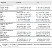
All of the hormones that were tested can be seen in this table:
1785949382989.webp

ME/CFS had lower baseline plasma DOPAC and DHPG before exercise, but not the next day. All other baseline hormones were normal.
CFS subjects had lower plasma levels of DHPG and DOPAC, but there were no differences between CFS and control subjects in the levels of these metabolites the next day (data not shown). There were no other differences in baseline hormone levels between the groups.
That's interesting, because the NIH deep phenotyping study found the same two metabolites were low, except they tested in the cerebrospinal fluid. However, Walitt et al. also found significantly lower DOPA, which this study did not (though it was nonsignificantly lower in this study).
Walitt et al. 2024 said:
In cerebrospinal fluid, the PI-ME/CFS group had statistically significant decreased levels of DOPA, DOPAC, and DHPG (Fig. 6a–c). Levels of norepinephrine, cys-DOPA, and dopamine did not differ between the groups. These results did not change after excluding data from participants taking central-acting medications.
 
Last edited:
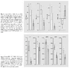
These figures show a subset of the hormone response measurements (4 minutes after exercise minus before exercise) for the two groups. It includes all those that were significantly different, plus a couple that were not:
1785949539059.webp

CFS subjects had lower plasma ACTH (F = 5.73 with 1, 25 d.f., p < 0.05), TSH (F = 4.99 with 1, 25 d.f., p < 0.05) and PRL (F = 4.94 with 1, 25 d.f., p < 0.05) responses than controls, and a trend toward greater GH responses (F = 4.17 with 1, 25 d.f., p = 0.08; fig. 1). Similar results were obtained in the analyses of GH responses when body mass index was included as a covariate. The plasma EPI response was also lower in CFS subjects than in controls (F = 7.80 with 1, 25 d.f., p < 0.01), but the NE response was not (F ! 1 with 1, 25 d.f., p 1 0.1; fig. 2). With maximum heart rate in the ANCOVA model, the groups did not differ significantly in their EPI responses because of a correlation between these variables (see below and fig. 3).

So there were significantly smaller changes after exercise in the ME/CFS group for ACTH, TSH, prolactin, epinephrine, DHPG, DOPA,and DOPAC, and an almost significantly larger growth hormone response.

As several participants in the ME/CFS group did not achieve maximum effort, the obvious question is whether that is the reason for smaller responses. Thankfully, they answered that question by re-analyzing with only those that achieved maximum effort. They got the same results, except the larger growth hormone response in ME/CFS was now significant.
These 12 CFS subjects achieved the same maximal heart rate, respiratory exchange ratio and blood lactate levels as control subjects. Reanalyses of the hormonal response data including only these 12 subjects in the CFS group produced the same results as the initial analyses. In the case of GH, a marginal effect (fig. 1) became a significant difference between CFS and control subjects
 
Last edited:
Interestingly, some of these findings were the opposite of what they would expect from people who are less athletic. (Primarily epinephrine and growth hormone responses.)
The diminished responses of CFS subjects to exercise, particularly the EPI response, was unexpected, given that people with greater levels of fitness have lower NE and EPI responses to exercise [17, 38]. Not only is this true in healthy subjects, but catecholamine responses to exercise also decrease after training and increased fitness in cardiac patients with ischemic heart disease [15]. In addition to changes in catecholamine responses, exercise training in healthy subjects will increase the GH response to exercise testing [17]. Given these findings, it might be expected that deconditioned CFS patients would have exaggerated NE and EPI responses to exercise and perhaps lower GH responses. However, our results indicate the contrary.
 
Back
Top Bottom