Preprint Initial findings from the DecodeME genome-wide association study of myalgic encephalomyelitis/chronic fatigue syndrome, 2025, DecodeMe Collaboration

The relevance here is that in the first exposure of the DecodeME data to the advisory board Chris showed a nice peak in MHC. I was puzzled when I saw the published data with a peak for BTN2A2 on chromosome 6 but nothing else. As far as I can see the reason is that BTN2A2 is within MHC.

Would it be an idea to ask Chris if this is the case? It seems like a very important point to get clear on.
 
To put this into a biological systems context, some physiology is required. In the gastrointestinal tract, epithelial cells proliferate and die rapidly. The division of these cells occurs at the base of villi, and cells are pushed upwards by subsequent divisions to the tip where they enter apoptosis and shed off into the lumen. Netrin-1 is produced in the base of the villi, so a gradient of netrin is present that is weakest at the tip. In normal physiology, the presence of netrin-1 inhibits DCC-mediated cell death until the epithelial cell reaches the tip of the villus, where the now unbound DCC causes the cell to enter apoptosis. In a cancer state, the absence of DCC prevents the gradient from having an effect on the cell, making it more likely to continue to survive.
DCC is a netrin-1 receptor, and it seems to be relevant to the functioning of epithelial cells of various sorts.

Predicted to be involved in several processes, including establishment or maintenance of epithelial cell apical/basal polarity
What is Epithelial Cell Polarity?
  • Asymmetrical Structure:
    Epithelial cells are oriented with distinct "poles" – an apical surface that faces a lumen or the outside of the organism, and a basolateral surface that faces other cells or the underlying extracellular matrix (basement membrane).

  • Structural Organization:
    This orientation allows the cells to form organized sheets and maintain their position within tissues.

  • Functional Barrier:
    Polarity is vital for the selective transport of ions, water, and other molecules across the epithelial layer, ensuring a controlled passage between different compartments of the body.
I thought epithelial polarity was an interesting idea linking those two genes, so just throwing it out there. Apparently bacteria can disrupt epithelial cell polarity, although I'm not sure how it would stay disrupted. I think T cells can disrupt epithelial cell polarity too, e.g.

Co-culture with acutely and chronically activated T cells decreased the magnitude of ion flux through the pore pathway, which was maintained in the presence of acutely activated T cells. Chronically activated T cells after 30 hours induced a precipitous increase in the magnitude of both ion and molecular flux, resulting in an increase in the unrestricted pathway, destruction of microvilli, expansion in cell surface area, and cell death. These fluctuations in permeability were the result of changes in the assembly and expression of tight junction proteins, cell morphology, and viability.


If epithelial cell polarity was disrupted, that might make problems related to the absorption of molecules across epithelium. For example, vascular epithelial issues could cause faulty fluid and ion homeostasis, maybe accounting for muscle weakness and pain.

Maybe the flow of water and ions across the faulty vascular epithelium would be okay when the body is at rest. But when more demand is placed on the body, maybe when there are more activated T cells, that would result in a faulty vascular epithelium not delivering what is needed to tissues (or taking waste away).
 
My layman understanding:
  • One allele, HLA-DQA1*05:01, was significantly associated with ME/CFS.
  • Its frequency was lower in cases (21.7%) compared to controls (23.2%) (the finding held even when they restricted analysis to a genetically more uniform group).
  • The association was very statistically strong (p = 1.4 × 10^{-10}).
However:
  • They checked related alleles (HLA-DRB1*03:01 and HLA-DQB1*02:01) but didn’t find strong associations (p-values were not significant: 0.27 and 0.042).
  • This was a bit surprising, since these alleles are often inherited together with HLA-DQA1*05:01.
Next steps:
They caution that the results are based on imputed data. They want to repeat the analysis using directly imputed HLA alleles for both cases and controls together to confirm whether this association is real or not.
 
it may also be worth remembering that 'MHC' originally refers to gene products that determine histocompatibility of allograft cells - which is not to do with their antigen presenting function.
Indeed. And the alleles that matter for compatibility are the ones in the actual HLA protein complexes.
 
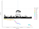
A gene that hasn't been disucssed much is TAOK3 on chromosome 12 (it wasn't a Tier 1 gene). It has been previously been associated with Lupus at around the same region as in DecodeME. The vertical dotted line in the graph below shows the location for the Lupus hit (12:118244946) with the SNP summary data from DecodeME.


1758826778973.png

The Lupus GWAS said this about it:
We also identified a missense variant in TAOK3 (the gene for tau kinase 3) as the top association signal in this locus. The risk allele (rs428073-T) substitutes the 47th amino acid of TAOK3 fromserine to asparagine (S47N), whose functional role remains unknown. S47 is located at the loop region between strands β2and β3, and the substitution should not change the overall strucure of the protein, despite being well conserved among orthologous proteins during evolutionary courses (SupplementaryFigure 10, on the Arthritis & Rheumatology website at https://onlinelibrary.wiley.com/doi/10.1002/art.42021). Taok3 plays animportant role in DNA damage–induced activation of the p38/MAPK14 stress-activated MAPK cascade. It enhances T cell receptor signaling by regulating its negative feedback by SH2domain–containing phosphatase 1 (44), and Taok3 deficiency in mice was found to cause defects in the development of marginalzone B cells but not follicular B cells (45).
 
"It enhances T cell receptor signaling by regulating its negative feedback by SH2domain–containing phosphatase 1 (44), and Taok3 deficiency in mice was found to cause defects in the development of marginalzone B cells but not follicular B cells (45)."

Intriguing. An influence on SHIP1 would make some sense. Marginal zone B cell behaviour is odd in lupus too.
 
A gene that hasn't been disucssed much is TAOK3 on chromosome 12 (it wasn't a Tier 1 gene). It has been previously been associated with Lupus at around the same region as in DecodeME. The vertical dotted line in the graph below shows the location for the Lupus hit (12:118244946) with the SNP summary data from DecodeME.


View attachment 28547

The Lupus GWAS said this about it:

Is this for the same hit that was annotated to SUDS3 in DecodeME?
 
Interesting, so it seems like this is another case where the final annotation for DecodeME was chosen based on high number of [edit: coloc] tissues. The top SNP is within an intronic region in DecodeME, and seems to be quite a long deletion. The mutation in the lupus paper you posted was in a protein-coding region which might explain stronger effect of the mutation.

Also, interesting that 3 proteins in that whole region that looks to be in LD have well-established links to MAPK signaling (SUDS3, TAOK3, and PEBP1)
 
Last edited:
I've copied @ME/CFS Science Blog's chart and some discussion about TAOK3 to the discussion thread we had on SUDS3, renaming it Chromosome 12 SUDS3, TAOK3. Probably those discussion threads should have been titled about the identified regions of interest rather than individual genes.

Genetics: Chromosome 12: SUDS3, TAOK3

Those charts with the DecodeME results and gene positions are really useful, ME/CFS Science Blog.
 
Those charts with the DecodeME results and gene positions are really useful, ME/CFS Science Blog.
Thanks I should probably mention that I zoom out a bit more (1Mb) than most tools like LocusZoom (around 250kb) to get an overview of the entire region around the hit. The implicated genes are probably closer to the top SNP then the region I show (so don't pay too much attention to the genes at the borders of my graphs).
 
Reading some GWAS in other illnesses made me appreciate DecodeME even more.

Other GWAS are usually based on (1) extracting data from big databases like the UK biobank, Finngen, AllofUS, 23andME etc. where the case definition was often poor or (2) on multiple cohorts that are combined into a single meta-analysis which likely has extra problems in terms of preventing confounding.

I haven't seen many GWAS where they collected the entire sample in one study like DecodeME did. This had the benefit that the case definition could be stricter defined using extra questionnaires and that the researchers could include various questions that might help their analysis. DecodeME was in contact with all its participants that donated DNA, which is often not the case for other GWAS.

I'm curious to see if some of the hits we see are due to a comorbidity such as depression or pain (if the signal is the same in ME/CFS patients without depression or without pain). Hoping that the questionnaire data will allow such an analysis.
 
Back
Top Bottom