Microvascular Dysfunction and Basal Membrane Thickening in Skeletal Muscle in ME/CFS and Post-COVID, 2025, Slaghekke et al

ScoutB about Rob Wust said:
He suggested this thicker capillary wall / reduced interior space could be messing with gas exchange into tissue and could be upstream of problems pwME have with muscles. I'm wondering, if this result turns out to be real in muscle, is it possible that the same thing could be wrong with capillaries in the brain?

I really hope that's not happening in the brain.
 
Last edited by a moderator:
I really hope that's not happening in the brain.
It certainly doesn't sound ideal. Am I right in thinking it would be hard to tell? Considering we are only able to find this in muscle by taking biopsies (rather than imagining).

On their EM findings, reduced capillaries, abnormal endothelial cells and basement membrane thickening / duplication I screen-grabbed in post #48 look to replicate findings (not universal in that n=11 cohort)
Slowly collecting my thoughts and writing a response to your reply in the other thread @SNT Gatchaman. In the meantime, I was so foggy when I first read this post I did not realize we now have two studies finding somewhat thickened basement membranes. Is it possible for someone to add the "capillary basement thickness" tag to this thread too? I assume I can't? idk how tagging works
 
Thanks for collecting all that together @SNT Gatchaman. Is it worth having a thread on this topic (on "capillary thickness" or more generally on the weird results showing up in these biopsies)? Has this been studied much in other diseases?

I might try to answer these questions myself when I'm less foggy. For now all I have is that wikipedia says
In histopathology, thickened basement membranes are found in several inflammatory diseases, such as lichen sclerosus, systemic lupus erythematosus or dermatomyositis in the skin, or collagenous colitis in the colon.
Btw be warned the lichen sclerosus wiki page is probably not safe for work unless you are like a gynecologist maybe.
 
I would take any bet that this is 1. a real and disease relevant phenomenon and 2. occurring in the ME/CFS brain.
For (1) I would like to see the correlation tested between such muscle biopsy findings and eg 1H and 31P MR Spectroscopy to Assess Muscle Mitochondrial Dysfunction in Long COVID (2024) with at rest and exercise challenge.

For (2) Maybe, but the brain microvasculature is very different, particularly in regard to the blood-brain barrier. It might be that the brain microcirculation is affected but in a different manner, possibly including via circulating factors generated from the muscle distress and the related immune response.

I think we've only had very limited post-mortems for (very) severe ME cases and I'm not aware of capillary thickness being reported — in muscle or brain.


Awaiting RECOVER results from Researching COVID to enhance recovery RECOVER tissue pathology study protocol: Rationale, objectives, and design (2024)
 
For (1) I would like to see the correlation tested between such muscle biopsy findings and eg 1H and 31P MR Spectroscopy to Assess Muscle Mitochondrial Dysfunction in Long COVID (2024) with at rest and exercise challenge.

For (2) Maybe, but the brain microvasculature is very different, particularly in regard to the blood-brain barrier. It might be that the brain microcirculation is affected but in a different manner, possibly including via circulating factors generated from the muscle distress and the related immune response.

I think we've only had very limited post-mortems for (very) severe ME cases and I'm not aware of capillary thickness being reported — in muscle or brain.


Awaiting RECOVER results from Researching COVID to enhance recovery RECOVER tissue pathology study protocol: Rationale, objectives, and design (2024)
Re 1, sounds interesting, I am quite confident I could get funding for such a project. So, if you had a team in mind, please reach out.

Re 2, I understand this (and other evidence, hamster models eg are very intersting) is very indirect, but it's good enough for me to make that bet.

 
Last edited:
if you had a team in mind

For a team, I'd start with the Wüst, van Vugt et al team in Amsterdam and see if they have collaborations available with local MR researchers. There may already be someone relevant involved in that team. I'd envisage an extension of their pre- / post- single CPET biopsy paradigm, augmented with pre- / post- MR spectroscopy (± NIRS).

Spit-balling. You'd probably want to do each MRI before each biopsy, 10 days apart as before. The MRI uses an exertion paradigm but is looking at calf muscles using an exercise band, whereas the biopsy is in thigh muscle (but you don't want a sore leg in the magnet, even if you biopsied the other side).

You can't exert the thigh muscles in the MRI because that would move the thigh out of the coil position (and there isn't space anyway). The CPET uses an upright cycle ergometer, which I expect works the thigh muscles much more than calf muscles. Perhaps you could also do MRI spectroscopy on resting thigh muscle to sample it as well, but that would add time on magnet. Apart from the CPET, doing 2 rounds of MRI and biopsy would be demanding on the participants though there would be time for rest between each component.

In addition, as that would only be looking at patients capable of CPET, I think it would be informative to at least biopsy a couple of moderate+ pwME at rest. That could potentially even be in their own home though of course an MRI can't travel to them and it's likely a more severe person wouldn't be able to tolerate an in-hospital MRI. Maybe passive NIRS could show something.

Alternatively, the radiology team that did the MR spectroscopy study (Oxford/Radcliffe team) might collaborate with local exercise physiologists, interventional radiology and pathology people for the biopsies. Depending on funding options, the Oxford and Amsterdam teams might even collaborate to make this multi-centre, though equally it could start with a single-centre pilot. With a collab potentially the patients could even be scanned and biopsied in the UK and the Amsterdam team could do their established tissue analysis. That would benefit from the established skills and techniques on the imaging and tissue assessment sides with each.

Just some ideas, but there are probably practical aspects I'm failing to consider. But to my mind muscle findings are worth pursuing at length, not least because, while we're working things out, the gold standard of direct tissue analysis following exertional trigger is available for muscle, where it isn't for brain. Advanced neuroimaging techniques are helpfully closing the gap though, eg DTI-ALPS, white matter microstructure diffusion imaging and myelin water fraction.
 
Last edited:
For a team, I'd start with the Wüst, van Vugt et al team in Amsterdam and see if they have collaborations available with local MR researchers. There may already be someone relevant involved in that team. I'd envisage an extension of their pre- / post- single CPET biopsy paradigm, augmented with pre- / post- MR spectroscopy (± NIRS).

Spit-balling. You'd probably want to do each MRI before each biopsy, 10 days apart as before. The MRI uses an exertion paradigm but is looking at calf muscles using an exercise band, whereas the biopsy is in thigh muscle (but you don't want a sore leg in the magnet, even if you biopsied the other side).

You can't exert the thigh muscles in the MRI because that would move the thigh out of the coil position (and there isn't space anyway). The CPET uses an upright cycle ergometer, which I expect works the thigh muscles much more than calf muscles. Perhaps you could also do MRI spectroscopy on resting thigh muscle to sample it as well, but that would add time on magnet. Apart from the CPET, doing 2 rounds of MRI and biopsy would be demanding on the participants though there would be time for rest between each component.

In addition, as that would only be looking at patients capable of CPET, I think it would be informative to at least biopsy a couple of moderate+ pwME at rest. That could potentially even be in their own home though of course an MRI can't travel to them and it's likely a more severe person wouldn't be able to tolerate an in-hospital MRI. Maybe passive NIRS could show something.

Alternatively, the radiology team that did the MR spectroscopy study (Oxford/Radcliffe team) might collaborate with local exercise physiologists, interventional radiology and pathology people for the biopsies. Depending on funding options, the Oxford and Amsterdam teams might even collaborate to make this multi-centre, though equally it could start with a single-centre pilot. With a collab potentially the patients could even be scanned and biopsied in the UK and the Amsterdam team could do their established tissue analysis. That would benefit from the established skills and techniques on the imaging and tissue assessment sides with each.

Just some ideas, but there are probably practical aspects I'm failing to consider. But to my mind muscle findings are worth pursuing at length, not least because, while we're working things out, the gold standard of direct tissue analysis following exertional trigger is available for muscle, where it isn't for brain. Advanced neuroimaging techniques are helpfully closing the gap though, eg DTI-ALPS, white matter microstructure diffusion imaging and myelin water fraction.

Thank you very much for your thoughtful suggestions. I would like to provide a brief but clearer overview of the current situation (here in Vienna).

The WE&ME Foundation is presently undergoing a restructuring process for the 'Johadamis Grant', which is valued at €100,000. The intention is to relaunch the grant next year with an updated framework.

Although the Foundation has not yet established direct contact with the Science4ME Committee, this is expected to occur, as the Foundation plans to include at least 50 percent patient representation in the jury responsible for evaluating proposals.

(The final funding decision will remain with the Foundation’s Board of Directors, but many procedural and structural details are still in development.)

For more context: I am a very severe ME patient and serve on the Foundation’s advisory board, but I am not in a position to handle all these organisational matters myself anymore (I was involved in allocating funding before).

Long story short, if you believe there is a clear ME/CFS-specific application for these funds (like you are outlining here), or if you have concrete ideas regarding how they could be effectively deployed, the Johadamis Grant module might be a good opportunity to push in that direction next year.

One of the bigger bottlenecks (apart from the lack of fundraising/funds - by far the biggest issue still) currently is actually bringing together applicants and charities - patients scouting for potentially worthy projects is currently underemployed and undervalued.

Thank you again for your engagement.
 
Last edited:
There seems to be some new info about this project on page 12 here https://www.amsterdamumc.org/download/ams-book-of-abstracts-2026

Welcome to the forum. Thanks, here is the page 12 abstract from the above document, which is a book of abstracts from the Amsterdam Movement Sciences research meeting on April 2, 2026:

Impaired Skeletal Muscle Oxygenation and Microvascular Dysfunction in Long Covid and ME/CFS
Anouk Slaghekke, Braeden T Charlton, Jelle Y Huijts, Brent Appelman 2, Ellen A Breedveld 1, Wendy Noort 1, Frank W Bloemers 3, Paul van Amstel 3, Jelle J Posthuma 4, Richie P Goulding 1, Michèle van Vugt 5, Rob C I Wϋst 1

1 Vrije Universiteit Amsterdam, Department of Behavioral and Movement Sciences, Amsterdam Movement Sciences, The Netherlands

2 Centre for Experimental and Molecular Medicine, Amsterdam UMC, The Netherlands

3 Department of Trauma Surgery, Amsterdam UMC, The Netherlands

4 Division of Surgery, Flevoziekenhuis, Almere, The Netherlands

5 Department of Internal Medicine, Division of Infectious Diseases, Amsterdam UMC, the Netherlands
Introduction
Long COVID and Myalgic Encephalomyelitis/Chronic Fatigue Syndrome (ME/CFS) are debilitating multisystem diseases characterized by brain fog, reduced exercise capacity, and post-exertional malaise. Ineffective matching of oxygen delivery to consumption in skeletal muscle can contribute to a lower exercise capacity and fatigue. Therefore, this study examines skeletal muscle oxygenation and possible alterations in skeletal muscle microvasculature.

Methods
Markers of skeletal muscle microcirculation were assessed in Long COVID (n=24), ME/CFS (pre-2020 diagnosis, n=26), and healthy controls (n=30). Participants underwent a maximal cycling exercise test for determination of maximal oxygen uptake (VO2max) with simultaneous muscle oxygenation assessment (near-infrared spectroscopy). Vastus lateralis muscle biopsies were analysed using immunohistology and electron microscopy to assess capillary ultrastructure, capillarization, and basal membrane thickness.

Results
Both patient groups obtained lower VO2max (p<0.0001) and peak power output (p<0.0001), despite no alterations in maximal heart rate or ventilatory equivalents. Tissue O2 uptake (p=0.001) and vasodilatory capacity (p=0.011) were lower in Long COVID and ME/CFS patients. Even though capillarization was only lower in ME/CFS patients (p<0.0005), both patient groups showed significantly lower capillary contact lengths (p<0.0001), primarily driven by decreased capillary tortuosity (p<0.0001).
Additionally, both patient groups displayed substantial capillary basement membrane thickening (p<0.0001), with a maximum of 62.7% in healthy controls and a minimum of 63.2% in patients.
Ultrastructural analysis further confirmed basement membrane thickening alongside multiple markers of endothelial dysfunction, including microvacuolization, endothelial hypertrophy, and signs of degeneration.

Conclusion
Microvascular impairments in patients with Long COVID and ME/CFS suggest inadequate skeletal muscle oxygenation, contributing to fatigue and post-exertional malaise. The pronounced basement membrane thickening may serve as a novel diagnostic biomarker and provide new insights into the pathophysiology of Long COVID and ME/CFS.
iMarkup_20260725_094934.webp
 
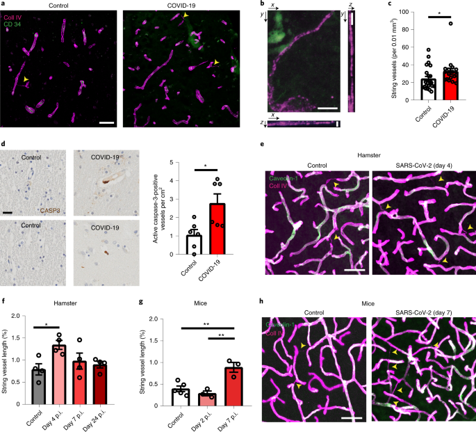
Is it possible that the basement membrane thickening observed here is a compensatory mechanism for increased type IV collagen degradation due to elevated MMP-9 observations in ME/CFS?
Perhaps this is just speculation with the weak evidence base thus far?
 
Is it possible that the basement membrane thickening observed here is a compensatory mechanism for increased type IV collagen degradation due to elevated MMP-9 observations in ME/CFS?
Perhaps this is just speculation with the weak evidence base thus far?

I think that is quite hard to argue. If the MM9 was doing anything the most obvious finding would be thinner BM. The endothelium might recognise that and replace it but what would explain making the BM thicker? My understanding is that MMPs in blood are generally switched off by inhibitors.
 
I suppose repeated endothelial cell damage/death leading to new basement membrane layers during replacement seems more likely (I am no expert, just speculating).

Except that what has been reported here does not look to me like any known pathological process seen in other conditions. Damage to capillaries tends to be very local, not generalised, except in things like amyloidosis. And generalised capillary change tend to be associated with increased permeability (paradoxically, despite the thickening) and no muscle oedema has been reported in ME/CFS. The difficulty I have with this report is that it claims to show something that, in the wider experience of pathology, ought to come with things that we don't see in ME/CFS. I am not even sure that it would make sense of fatigue or weakness.
 
Back
Top Bottom