Trial Report Plasma cell targeting with the anti-CD38 antibody daratumumab in ME/CFS -a clinical pilot study, 2025, Fluge et al


Antibodies to peptides are pretty meaningless. They seem to have found antibodies that bind to peptides from a range of proteins that share some sequence similarity to microbial proteins, which is almost certainly completely irrelevant. Peptides with similar sequences exist everywhere in everything. If they had found a qualitative difference in antibodies to one protein I would have been interested. This sort of result has been produced for all sorts of diseases for decades and tells us nothing as far as I can see.
 
Here is the revised PILOT protocol.

“Protocol ver. 1.6: Amended treatment protocol; this will apply to four additional patients included in Q1 2025, as well as retreatment of one previously included patient with partial relapse. Patients will receive the first daratumumab subcutaneous injection at week 0. For patients with signs of clinical response after the first injection, a second injection will be given at 24 weeks, and a third injection at 48 weeks.

At week 10, if there are no signs of a clinical response, a second daratumumab injection will be given, and, subject to a clinical response, the third and fourth injection will be given at week 24 and 48. If patient experiences no response after the second injection, no further daratumumab will be given.”

Can be found here.


I think this jibes with a comment @Jonathan Edwards made earlier somewhere on this forum that one initial dose would be sufficient to deplete long-lived plasma cells.
 
I’m assuming that they didn’t go with this revised protocol for the full Phase 2 study because they didn’t want to take the risk that weren’t fully depleting LLPC’s.

With this very small sub-sample they could take the risk of doing fewer injections.

FWIW, this is very different from the multiple myeloma dosing, which I believes entails 8 initial injections.
 
Antibodies to peptides are pretty meaningless. They seem to have found antibodies that bind to peptides from a range of proteins that share some sequence similarity to microbial proteins, which is almost certainly completely irrelevant. Peptides with similar sequences exist everywhere in everything. If they had found a qualitative difference in antibodies to one protein I would have been interested. This sort of result has been produced for all sorts of diseases for decades and tells us nothing as far as I can see.
Is this way better for finding autoantibodies? cell assay?
Autoimmune Basis for Postural Tachycardia Syndrome
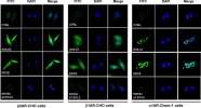
We used receptor‐transfected cells to demonstrate specific autoantibody binding to the receptors. Sera from a POTS subject who demonstrated activating autoantibodies to β1AR, β2AR, and α1AR, sera from a healthy control subject and rabbit polyclonal antibodies to each receptor as positive controls were separately incubated with these cells without and with an excess of their respective ECL2 target peptide. These images are shown in Figure 6. There was significant binding to each of the receptors from the POTS sera, which was completely blocked by preabsorption with the specific ECL2 peptides. No significant binding to the cells from the control sera was observed.

jah3-3-e000755-g6.jpg
 
Immunofluorescence assays like the pictures shown have been the gold standard for pathogenic autoantibodies. They correlate robustly if not universally with diseases we know to be antibody driven. Some would say you need to stain live cells but I don't think that is necessary. Functional assays as described here ought to be good but there are quality control and batch control problems that make it very difficult to assess their reliability in reports like this.

So, yes, the staining assay is a good one. But in Scheibenbogen's studies the rate of positives in MECFS patients is barely different from normal. Which means that the antibodies cannot possibly be directly responsible for pathology. It is said that many people with MECFS have "POTS" but if so this MECFS POTS isn't due to these GPR antibodies. Even for a non MECFS cohort of POTS like this paper it is pretty extraordinary that they were all positive and controls all negative. In other words this looks a bit too good to be true. And the trouble is that we repeatedly see invalid study designs for things like this - like only including people in your cohort you know to be positive beforehand.

I think it is quite possible that at subset of people diagnosed with POT have an autoantibody mediated problem but the evidence is that this is not generally relevant to MECFS.
Nice
I didn't ask this in the context of the disease but more in the direction of focusing on the assay type.
Thanks
 
Immunofluorescence assays like the pictures shown have been the gold standard for pathogenic autoantibodies. They correlate robustly if not universally with diseases we know to be antibody driven. Some would say you need to stain live cells but I don't think that is necessary. Functional assays as described here ought to be good but there are quality control and batch control problems that make it very difficult to assess their reliability in reports like this.

So, yes, the staining assay is a good one. But in Scheibenbogen's studies the rate of positives in MECFS patients is barely different from normal. Which means that the antibodies cannot possibly be directly responsible for pathology. It is said that many people with MECFS have "POTS" but if so this MECFS POTS isn't due to these GPR antibodies. Even for a non MECFS cohort of POTS like this paper it is pretty extraordinary that they were all positive and controls all negative. In other words this looks a bit too good to be true. And the trouble is that we repeatedly see invalid study designs for things like this - like only including people in your cohort you know to be positive beforehand.

I think it is quite possible that at subset of people diagnosed with POT have an autoantibody mediated problem but the evidence is that this is not generally relevant to MECFS.
Another question is what type of IgG is binding to the receptor?
IgG4 is not known to bind to receptors like IgG1/3 does.
 
IgG4 does not bind to IgGFc receptors via its Fc but it can bind to any sort of receptor (for any other protein) via its Fab antigen binding end.
Is there a way to isolate the IgG groups from the serum and then to check them individually like they did in the study?
Also, if the IgG binds through Fc, where is the inflammation/damage?
 
Back
Top Bottom