View attachment 34487
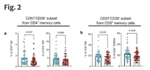
Just to situate the interpretation of this paper, these are the figures that the claim of possible indirect evidence of chronic antigen stimulation are drawing from. Each panel is missing one T cell subset that would have been tested (likely not shown because of lack of group differences). CD28-CD57- is also a weird population to zoom in on—the one typically associated with chronic antigen is CD28-CD57+, since CD57 increases as CD28 decreases.
The text claims that CD28-CD57- represents an “early senescence” phenotype, but found no difference in the classic “senescence” phenotype. Which doesnt particularly fit with a story of chronic antigen stimulation. These are long-lived T cell populations. If this is a real difference and not a chance artifact, why would only the small subpopulation of a subpopulation of cells just barely starting to reflect signs of chronic stimulation show a difference in number? We also don’t know the antigen specificity of these subsets: a virus-specific population of these T cells would only skew overall population numbers if they were a significant proportion of the memory T cells, which would happen downstream of clonal expansion, which we don’t have strong evidence of in ME/CFS.
When you look through the whole text, figures, and supplemental figures you get a sense of the sheer scale of different subpopulations measured and compared between groups. What isnt shown in this figure is the comparisons across CD4 and CD8 CMs, EMs, and TEMRAs for all the other combinations of CD57 and CD28. Which is a very large number of things to check with no p-value correction for multiple testing. And that’s just for one figure.
Even if these findings are real and not the result of random chance (being extremely generous I might say they somewhat fit with the “increased markers of exhaustion” line from the Grimson lab) this is not strong evidence of chronic antigen stimulation from a persistent virus. These T cell markers respond to all kinds of things, including other transient immune signaling that has nothing to do with a hidden virus.
And no, none of this is evidence of a “hyperinflammatory” or even an “inflammatory” state. You can’t define that from T cell populations alone.