
[Abstract] Immediate Plasma Lipid Perturbations Following Cardiopulmonary Exercise Testing In [ME/CFS], 2026, Christopoulos, Armstrong et al

forestglip

Administrator
Staff member
Immediate Plasma Lipid Perturbations Following Cardiopulmonary Exercise Testing In Myalgic Encephalomyelitis/ Chronic Fatigue Syndrome (ME/CFS)

Christopoulos, Elena M.; Huang, Katherine; Howden, Erin J.; Talbot, Jack S.; Armstrong, Christopher W.; Moneghetti, Kegan J.

PURPOSE
Previous research has shown that myalgic encephalomyelitis/chronic fatigue syndrome (ME/CFS) patients experience post exertional malaise (PEM) after performing a cardiopulmonary exercise test (CPET). However, it is currently unknown if there are metabolic perturbations that appear after exertion that suggest a patient may experience PEM or predict PEM severity. Our aim was to characterise the lipidomic alterations that occur following exertion in ME/CFS in comparison with healthy controls.

METHODS
Twenty-five participants with ME/CFS and 25 healthy controls (mean age 44.1 ± 12.7 and 43.3 ± 13.2 years, 92% females respectively) completed a maximal exertion incremental ramp protocol CPET. Blood was collected at baseline (prior to exertion) and at 2 and 30 minutes following the CPET and was centrifuged to obtain plasma.

Liquid chromatography mass spectrometry was used to perform targeted lipidomics, with 794 predefined targets in the metabolite database, to determine the relative abundance of the lipids. Group differences in lipid abundance were assessed using either Student’s or Welch’s t-tests and Mann-Whitney U tests as appropriate.

RESULTS
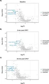
Reduced relative lipid abundance was observed between the ME/CFS and control groups at baseline and at both 2 and 30 minutes following the CPET with 101, 60 and 39 significant differences respectively.

Additionally, consistent downregulation of lipids was identified in the ME/CFS group compared to controls A) at baseline, B) 2 minutes after VO2 peak, and C) 30 minutes after VO2 peak (Figure 1), with four lipids at each timepoint reaching statistical significance (FC threshold >1, q- value <0.05).

CONCLUSIONS
Patients with ME/CFS experience a baseline reduction in lipid metabolism and following exertion. This may indicate a delay in the increase in aerobic metabolism following exertion, indicating impaired post exertional metabolism recovery and persistent metabolic inefficiency underlying PEM.
1790252143399.webp

Web | DOI | PDF | Medicine & Science in Sports & Exercise | Abstract
 
Interesting to see low cholesteryl-esters. This could be due to reduced LCAT activity as it converts free cholesterol to cholesteryl esters on HDL.

They also found low PC, which is interesting because PC is used as an acyl donor for LCAT.

Also oxidative stress reduces LCAT activity as well, and oxidative stress found to be higher in ME/CFS.

Low LCAT activity causes reduced red blood cell deformability, which Ron Davis has found happening in ME/CFS patients.

I made a video about how oxLDL could cause reduced LCAT activity and lead to lower conversion of free cholesterol to cholesteryl-esters-


Both @DMissa and @MelbME have found low PC now
 
Last edited:
Both @DMissa and @MelbME have found low PC now
specifically lyso PC species in both studies - the one in the last study was the one that was discriminated all but one mild ME/CFS sample as well (per the dot plot of just that lipid's levels - no stats magic)

caveat is that last study is in cultured immortalised B cells and this one is in plasma

mechanistically I am not sure where this clue points us but it I am pleasantly surprised to see this pop up twice from studies done in the same city with same clinical criteria and so it may be a useful lead. I was more skeptical of seeing this only once and in cells that are quite removed from an in vivo context. I guess my first question would be: if there is a systemic difference in lipid handling what would mediate this? I still think this is less likely than a tissue-specific effect but perhaps there is some signal that pushes things the same way in circulating cells as it may in liver? (trying to think of where a plasma signature like this would be most likely to come from)

The chances of this occurring twice by chance are pretty low given the small number of significantly changed features that qualify for this comparison in both studies and given how many lipid classes and subclasses there are. To be clear: 3/5 significant features from the pooled results of both studies are reduced levels of Lyso PC lipids, out of several dozen categories of lipid that will have been detected in both. If this proportion was tested statistically you'd have a tiny p value. So I'm not yet sure what it might actually reflect but this concordance doesn't seem to be due to random chance. It is very unlikely.
 
Last edited:
specifically lyso PC species in both studies - the one in the last study was the one that was discriminated all but one mild ME/CFS sample as well (per the dot plot of just that lipid's levels - no stats magic)

caveat is that last study is in cultured immortalised B cells and this one is in plasma

mechanistically I am not sure where this clue points us but it I am pleasantly surprised to see this pop up twice from studies done in the same city with same clinical criteria and so it may be a useful lead. I was more skeptical of seeing this only once and in cells that are quite removed from an in vivo context. I guess my first question would be: if there is a systemic difference in lipid handling what would mediate this? I still think this is less likely than a tissue-specific effect but perhaps there is some signal that pushes things the same way in circulating cells as it may in liver? (trying to think of where a plasma signature like this would be most likely to come from)

The chances of this occurring twice by chance are pretty low given the small number of significantly changed features that qualify for this comparison in both studies and given how many lipid classes and subclasses there are. To be clear: 3/5 significant features from the pooled results of both studies are reduced levels of Lyso PC lipids, out of several dozen categories of lipid that will have been detected in both. If this proportion was tested statistically you'd have a tiny p value. So I'm not yet sure what it might actually reflect but this concordance doesn't seem to be due to random chance. It is very unlikely.
Can you please explain what lyso PC are? And just PC in general. Maybe in a brain fog friendly format. I'm too tired to look this up.
 
I posted a review article on lysophospholipids more generally at Lysophospholipid Mediators in Health and Disease (2022)

Phospholipids, the main component of biological membranes, usually have two fatty acid chains. Phospholipids with a single fatty acid are called lysophospholipids, even though their concentrations are much lower than those of conventional phospholipids in cells and tissues. Like phospholipids, lysophospholipids are classified by their polar head structure. For example, lysophosphatidylcholine (LPC) has choline in its polar head and lysophosphatidylserine (LysoPS) has l-serine. Lysophospholipids are also classified as glycerolysophospholipids or sphingolysophospholipids depending on whether they have a glycerol or sphingosine backbone, respectively.

We could perhaps summarise things on that thread.
 
@DMissa do you happen to know off the top of your head if there have been other plasma lipidomics studies that would have picked up a shift like this, if it were in their sample? Just trying to calibrate on how much this has been looked at before.
 
@DMissa do you happen to know off the top of your head if there have been other plasma lipidomics studies that would have picked up a shift like this, if it were in their sample? Just trying to calibrate on how much this has been looked at before.
It's tricky to interpret prior studies without any cells or tissue to compare to and they are pretty much always only on plasma/serum so no dice. Lipids in plasma are really affected by many lifestyle factors on top of many potential biological factors. Comparing this stuff across countries is probably futile imo.

I compare these two studies (one on plasma and one on cells) because they have been done in the same city at a similar time and with the same criteria (and probably some of the same people who tend to put their hands up for research). I think the post challenge measurements are interesting.. most of the prior studies of this sort haven't done this.

So there are a few axes of comparison possible here.

The problem with the lipid stuff is that a lot of different people have done it in a first pass sort of way using mass spec on plasma and then you never hear anything again. So we have a lot of early stage data that is hard to compare between different studies, (the individual studies themselves aren't giving particularly deep insights a lot of the time). This is why I am excited by the concordance here, because the inter-study comparison is less tenuous and there have been different, complementary things being done (more layers than usual). Altogether it could be the biggest step we've made in this area. Next we need an integrated medium sized study to tackle it from multiple angles to prove what the useful leads may or may not be in a very carefully selected cohort to get around the typical confounders (eg: aggressive medication exclusion criteria should be used) and to approach with focused hypotheses about specific mechanisms that are well reasoned to have a potential role in a plausible disease process
 
Last edited:
Ah I see, that makes sense. Thanks, very interesting!
I should clarify; I mean comparing these big datasets of hundreds or thousands of features that vary with lifestyle etc is the tricky bit. If a specific feature is identified as being of interest and if it is really part of a disease process it should probably be measurable in different places.
 
mechanistically I am not sure where this clue points us but it I am pleasantly surprised to see this pop up twice from studies done in the same city with same clinical criteria and so it may be a useful lead.

Just making sure , the people selected have been chosen so that they are not taking part twice correct ? studies done in the same city raises this risk
 
Back
Top Bottom