Deep Sequencing of BCR Heavy Chain Repertoires in Myalgic Encephalomyelitis/Chronic Fatigue Syndrome, 2025, Ryback et al

Do you mind listing what those 3 studies were? The first thing that comes to mind to check is whether all of them were done after Covid started.

This thread's study, plus the following two:
Plasma proteomic profiling suggests an association between antigen driven clonal B cell expansion and ME/CFS, 2020, Lipkin et al
IGHV3-23/30 and immunoglobulin kappa variable region 3–11 were significantly associated with ME/CFS without sr-IBS.

Skewing of the B cell receptor repertoire in myalgic encephalomyelitis/chronic fatigue syndrome, 2021, Sato et al
By applying a next-generation sequencing to determine the clone-based IGHV/IGHD/IGHJ repertoires, we revealed a biased usage of several IGHV genes in peripheral blood B cells from ME/CFS patients. Results of receiver operating characteristic (ROC) analysis further indicated a possibility of distinguishing patients from healthy controls, based on the skewed B cell repertoire. Meanwhile, B cell clones using IGHV3-30 and IGHV3-30-3 genes were more frequent in patients with an obvious infection-related episode at onset, and correlated to expression levels of interferon response genes in plasmablasts. Collectively, these results imply that B cell responses in ME/CFS are directed against an infectious agents or priming antigens induced before disease onset.

The first one isn't exactly the same as the other two since they couldn't discriminate between IGHV3-23 and IGHV3-30, and the ME/CFS group seemed to have individuals with both very high and very low amounts of the combination of these gene segments:
Milivojevic et al said:
Whilst 12 patients had extremely high levels (>100,000) of IGHV3-23/30, only 3 patients had extremely low levels (<25,000) of this protein.

I'm going to have to reread that one. I think I had misread before as it being IGHV3-23/30 on the BCR, but looks like it's actually antibodies in the first paper, with the following two being BCRs.

Here's the Ryback et al paper referencing the previous two studies:
BCR repertoire sequencing in ME/CFS and healthy controls (HCs) previously identified increased use of several Immunoglobulin Heavy Chain V (IGHV) gene segments in ME/CFS patients: IGHV-3-30 and IGHV3-30-3 gene usage was elevated in ME/CFS, particularly in patients who reported an infectious onset to their illness (19). The authors suggest this IGHV gene signature may represent a shared infection-elicited response among those ME/CFS patients.
This result putatively fits a signature of dysregulated IGHV3-30/IGHV3-23 antibody detected by plasma proteomic profiling (20).

With regard to COVID timing - I'm not sure about the Lipkin paper, though it was published near the start of the pandemic (2020), so the sample collection might have been before it. Sato et al says this:
Cohort 1 comprised patients recruited to NCNP hospital from October 2015 to December 2016. [...] To validate the results of BCR repertoire analysis, new patients with ME/CFS were recruited from NCNP hospital from January 2017 to November 2019, which comprised cohort 2

Ryback et al samples were from the CureME biobank, but I don't see dates of collection.
 
Last edited:
This thread's study, plus the following two:


The first one isn't exactly the same as the other two since they couldn't discriminate between IGHV3-23 and IGHV3-30, and the ME/CFS group seemed to have individuals with both very high and very low amounts of the combination of these gene segments:


I'm going to have to reread that one. I think I had misread before as it being IGHV3-23/30 on the BCR, but looks like it's actually antibodies in the first paper, with the following two being BCRs.

Here's the Ryback et al paper referencing the previous two studies:


With regard to COVID timing - I'm not sure about the Lipkin paper, though it was published near the start of the pandemic (2020), so the sample collection might have been before it. Sato et al says this:


Ryback et al samples were from the CureME biobank, but I don't see dates of collection.
Thanks! The conclusion from Sato et al. is exactly what I was going to say—none of these results look particularly like the BCR repertoires I’ve seen from a known auto-antibody mediated disease. What they do indicate to me is slightly differential efficacy of long term B cell response to vaccination or a pathogen encountered in the last few years. IGHV3-30 comes up specifically for SARS-CoV-2 spike protein, and since it is one of the more commonly used genes, I would imagine it could come up for seasonal viruses as well. Which might still point us towards underlying immune pathways in ME/CFS that regulate B cell responses, but a bit non-specifically at that.
 
Last edited:
IGHV3-30 comes up specifically for SARS-CoV-2 spike protein, and since it is one of the more commonly used genes, I would imagine it could come up for seasonal viruses as well.

Eg Longitudinal Multi-omics Analyses Identify Responses of Megakaryocytes, Erythroid Cells, and Plasmablasts as Hallmarks of Severe COVID-19 (2020) —

Analysis of immunoglobulin heavy-chain variable region (IGHV) family subunits showed a preponderance of specific V regions in COVID-19 patients compared with in controls, e.g., IGHV3-30 and IGHV3-23 were overrepresented in PBs and neutrophil-like cells during disease. In summary, we observed an increase in B cell clonality in COVID-19, and there was an increase of memory B cells and PBs, dominated by the IgA and IgG isotypes and a skewed use of the IGHV gene early during the disease course.
 
I'm not sure about the Lipkin paper, though it was published near the start of the pandemic (2020)

Noting that they do say the exact date in Milivojevik/Lipkin 2020:
Blood samples were collected into BD VacutainerTM Cell Preparation Tubes (CPT) with sodium citrate anticoagulant between June and October 2014, and centrifuged to pellet red blood cells.
 
Ryback et al samples were from the CureME biobank, but I don't see dates of collection.

I remember that 2019/20 winter before covid they were collecting but obviously stopped, as I had been scheduled to give more blood. I’m not sure exactly when they restarted but Caroline was doing limited home visits later in 2021 IIRC.

The Ryback paper mentions it uses the same samples as Dibble et al, published in Jan 2024 but that doesn’t mention dates either.

Would there have been time for the Biobank to collect, process, distribute and the team do their processing and research and write it up all in that window? Would there have been enough samples collected? And presumably unless there’s other requirements the Biobank get the older samples out into use first?

So I’d guess pre covid samples are most likely.
 
It's been almost a year and a half since this paper replicating high IGHV3-30 was published. 5 and 6 years since the other two papers reported high IGHV3-30.

If I was in charge of a bunch of ME/CFS money, one of my top priorities would be immediately:
  1. verifying this is not a fluke with more and larger studies.
  2. verifying that this is not something seen just because people with ME/CFS recently had infections or are more prone to infections, and is not a result of low physical activity.
  3. digging into the surrounding biology, as recommended by the authors: e.g. looking at expression of this gene in different isotypes of B cells.
Maybe it's my bad memory or I just don't read enough other studies, but it seems very rare that such a specific biological finding is seen in not only two but three studies, so this seems like some of the best low-hanging fruit we have.

I do think there's a good chance that even if it's a real finding, it's not interesting. For example, maybe people who are more prone to infections are more likely to get ME/CFS, and are also more likely to have high IGHV3-30. And it's possible it's not even a real finding, since only the mild/moderate participants in this study had high IGHV3-30. Maybe these are the reasons there doesn't seem to be a rush to study it more? Maybe there is a rush, but research just takes that long?
My sense is that there is some skepticism around B cell work due to small effect sizes and things like ritux trial failure. From some people, not all. But I do think people get hung up on specific corners of things (and make heuristic associations with words being good or bad), when in reality something like immune function is very interconnected and may have relevant steps going awry in different ways in different cell populations and to varying degrees of subtlety.

As someone moderately interested in whether B cells play a role, they will be included in an upcoming proposal but one that's asking different questions based on prelim data that strikes me as being more unusual and potentially impactful. If I could wave a wand to fund and follow up the work published in this thread without derailing this effort I would.

Don't know whether this provides useful insight following your question.
 
Last edited:
Germline based SARS-CoV-2 specific B cell repertoire motif identified with novel sequence based bioinformatic pipeline
Fridman, Daniel; Israitel, Lena; Shtewe, Areen; Hershberg, Uri
From abstract:
Utilizing data from an anti-SARS-CoV-2 vaccination study, we analyzed two properties of SARS-CoV-2 specific memory B-cells and compared them to the background immune repertoire.
Furthermore, we observed that SARS-CoV-2-specific B-cells show an increased tendency to utilize and conserve a specific CDR2 motif derived from the VH3–30 gene and its alleles.

Web | DOI | PMC | PDF | Frontiers in Immunology | 2026 | Open Access
 
As someone moderately interested in whether B cells play a role, they will be included in an upcoming proposal but one that's asking different questions based on prelim data that strikes me as being more unusual and potentially impactful
it's amazing how much changes in a month or two, but i've shifted future focus to T cells and finding ways to look at CNS based on weight of evidence atm. wanted to set record straight - not writing a b cell grant atm
 
Last edited:
it's amazing how much changes in a month or two, but i've shifted future focus to T cells and finding ways to look at CNS based on weight of evidence atm. wanted to set record straight - not writing a b cell grant atm
Does this mean you are no longer interested in the role of B cells or that looking at other routes may be more bang for the buck targets?
 
Knockdown of the long isoform of the prolactin receptor selectively targets pathogenic immune cells in systemic lupus erythematosus and averts glomerular pathology, 2026, Hamane et al

Hamane, Kory; Hu, Zunsong; Lima-Junior, Joao Rodrigues; Khani, Adeleh Taghi; Kumar, Anil; Ortiz, Ashly Sanchez; Guo, Xinying; Jung, Da Hae; Qin, Hanjun; Tao, Shu; Sanz, Ignacio; Meffre, Eric; Marzan, Katherine; Koff, Jean L.; Lorenson, Mary Y.; Gu, Zhaohui; Wu, Xiwei; Walker, Ameae M.; Swaminathan, Srividya

Abstract
Abstract Systemic lupus erythematosus (SLE) is an autoimmune disease characterized by chronic inflammation in multiple organ systems. While a clinical association between elevated levels of the hormone/cytokine, prolactin (PRL), and exacerbation of SLE has been recognized for some time, little is known about the mechanisms through which PRL affects the course of this disease. Here, we show that immune cells in SLE have aberrant splicing of the prolactin receptor (PRLR) such that the ratio of the long to short splice variants is increased. To determine whether the change in PRLR isoform expression was causal in this disease, we used a splice-modulating oligomer (SMO) that knocked down expression of the long splice variant (LFPRLR). Using patient samples ex vivo , SLE-prone mice in vivo , high-dimensional flow cytometry and single-cell RNA-sequencing, we demonstrate that the aberrant PRLR isoform expression in SLE both directly and indirectly drives production of autoreactive immune cell phenotypes. Thus, LFPRLR knockdown decreased the expression of type I interferon signaling/response genes known to be biomarkers and hub genes for SLE, reduced immunoglobulins with signatures considered autoreactive, and averted glomerular kidney damage in SLE-prone mice. Importantly, LFPRLR knockdown reduced pathogenic B cells and other immune subsets that drive B-cell activation, without negative impact on healthy donor counterparts. Current treatments for SLE adversely affect healthy cells and do not concurrently eradicate multiple pathogenic immune subsets. Since LFPRLR SMO does not share these disadvantages, knockdown of the LFPRLR represents a potential treatment strategy for SLE that merits further investigation. Graphical Abstract The LFPRLR represents an attractive therapeutic target in SLE. ( Left ) In addition to increased pituitary/circulating PRL, individuals with SLE exhibit aberrant increases in the production of autocrine/paracrine PRL by immune cells, and in their expression of specifically the long isoform (LF) of the PRLR. ( Middle ) Expression of the LFPRLR specifically enhances autoreactive immunophenotypes and promotes lupus nephritis. ( Right ) A splice modulating oligomer (SMO), that prevents synthesis of only the LFPRLR but not the short PRLR isoforms, reduces pathogenic immunophenotypes without affecting the normal counterparts of immune cells.

Web | DOI | PMC | PDF | bioRxiv

A bit over my head so I may be jumping a few steps as I tend to but maybe those with a better grip can chime in but is it fair to say this paper shows that prolactin can bypass some checkpointing in B cell maturation and selection which can lead to changes in b cell repertoire?

So we could potentially see a situation where transitory elevated or spiky prolactin levels could lead to elevated IGHV3-30? A downstream effect of a downstream effect potentially?
 
So we could potentially see a situation where transitory elevated or spiky prolactin levels could lead to elevated IGHV3-30? A downstream effect of a downstream effect potentially?

I suspect the connection is too tenuous to be worth investing in.

Most of this study is in 'SLE-prone mice', that have SLE for different reasons from humans with SLE. I tend to be very cautious about interpreting such things.
 
I suspect the connection is too tenuous to be worth investing in.

Most of this study is in 'SLE-prone mice', that have SLE for different reasons from humans with SLE. I tend to be very cautious about interpreting such things.
Thanks. Yes am always cautious of the ‘mouse with a condition’ angle.

There seem to be a few papers around the idea of long-form Prolactin Receptor (LFPRLR) knockdown mice and changes in immunoglobulin G (IgG) heavy chain and specific b cell populations.

The underlying broad idea seems to have started with a paper in 2002 called “Prolactin modulates the naive B cell repertoire, 2003, Peeva et al“. Maybe we’re just missing some specifics? Seems worth thinking about at least.

Papers re-posted here to not clog up this thread. But a few quotes below

Prolactin is a peptide hormone produced by the anterior pituitary gland that is critical in lactation. Prolactin can also be produced by lymphocytes, and both B and T cells express prolactin receptors. These findings have suggested that prolactin has immunomodulatory functions.

The DNA-reactive B cells had a follicular cell phenotype. Anti-DNA hybridomas demonstrated that prolactin alters selection of the naive B cell repertoire. The expansion and activation of anti-DNA B cells in prolactin-treated R4A-γ2b BALB/c mice was dependent on the presence of CD4+ T cells.

Finally, treatment with prolactin was unable to break tolerance in R4A-γ2b transgenic C57Bl/6 mice, suggesting that responsiveness of the immune system to prolactin is genetically determined.
 
Last edited:
Referring to some of the ideas in the papers I posted in the other thread

AIUI immune cells, particularly b cells (which we’ll focus on here, but also t cells and macrophages amongst others) can produce prolactin themselves as well as having prolactin receptors. These receptors look identical outside the cell but differ inside in their makeup (dictated by alternative splicing into long and short forms) and their effects. One form triggers the JAK/STAT pathway and increases expression of interferon stimulated genes and type I Interferon in the local environment. Then other doesn’t and so the balance of the two forms in a cell seems key.

There seems to be some potentially fun mechanisms here for localised prolactin and localised interferon too (paging @jnmaciuch )

This route to bypass checkpointing though seems like a potential route to a B cell repertoire shift like we saw in Audrey’s paper.

So a question for Audrey and @chillier (and others).

Can we re-examine the fastq data to look at CDR3 loop length and see of there are changes similar to those discussed in those papers?

Or look at skewing of the ratio of IgM to IgG specifically in the sub population of IGHV3-30 cells?

Are there other factors we can look at in this existing data to see if there are signs of a (potentially prolactin driven) shift or bypassing of checkpointing happening here?

I don’t know if it’s worth it or possible but using data we already have always seems like an attractive idea to me!
 
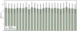
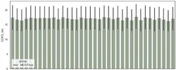
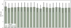
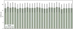
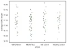
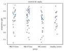
Audrey has quickly made these:
Wow thank you! I was more musing if this was possible or useful than expecting an answer. Very grateful and I guess clearly closes off that line of thought!

The fastqs should be publicly available
Looks like learning how to use them is on my todo list then :)
 
Last edited:
Thinking more about what this means and what it may tell us or rather what other questions to ask (of the data or the world in general).

I think we’ve already covered these in the thread before though

- Is this shift only showing up in mild/moderate as they have been active enough to trigger the underlying cause of it recently?

- Is the shift a sign of (an underlying genetic) protective factor which therefore shows up more in people who are mild/moderate?

What other possibilities do people think are there here?

I’m wondering if we can start to answer the second by looking at the data we already have. Or if not what other questions we can ask of it. Open to suggestions and ideas!

So far I’m looking at

And wondered if this would be useful
 
Last edited:
Back
Top Bottom