Deficient TRPM3-linked mitochondrial Ca2+ influx in natural killer cells associated with [ME/CFS], 2026, Magawa et al

forestglip

Moderator
Staff member
Deficient TRPM3-linked mitochondrial Ca2+ influx in natural killer cells associated with myalgic encephalomyelitis/chronic fatigue syndrome

Magawa, Chandi Tabeth; Eaton-Fitch, Natalie; Muraki, Katsuhiko; Marshall-Gradisnik, Sonya

Introduction
Myalgic encephalomyelitis/chronic fatigue syndrome (ME/CFS) is a multisystemic illness, commonly associated with dysregulation of the immune system including reduced cytotoxicity of natural killer (NK) cells and post-exertional neuroimmune exhaustion. Previously, transient receptor potential melastatin 3 (TRPM3) ion channel impairment associated with reduced Ca2+ mobilisation in NK cells from ME/CFS patients was reported.

To further explore the pathomechanisms involved in ME/CFS, we investigated the downstream impact of TRPM3 ion channel dysfunction on mitochondrial Ca2+ mobilisation in NK cells.

Method
Fluorescence live-cell imaging was used to investigate Ca2+ mobilisation in NK cells of (N = 10) ME/CFS, classified using Canadian Consensus Criteria, and (N = 10) healthy control (HC) participants. Cytoplasmic and mitochondrial Ca2+ entry was determined using Fluo-8 AM and Rhod-2 AM Ca2+ indicators, respectively.

The effect of TRPM3 modulation on Ca2+ mobilisation ex vivo, was examined using pregnenolone sulfate and ononetin to activate and inhibit the channel, respectively.

Results
Cytosolic Ca2+ influx amplitude and slope were significantly reduced (p < 0.001), with a significantly shorter T1/2 response (p = 0.001) in ME/CFS compared to HC. Ca2+ influx amplitude (p < 0.001) and slope (p < 0.041) into the mitochondria were significantly higher in ME/CFS compared to HC.

TRPM3 activation triggered pronounced cytosolic response (P < 0.001) accompanied by mitochondrial Ca2+ increase in HC. TRPM3-dependent cytosolic and mitochondrial Ca2+ mobilisation (P < 0.015) were significantly reduced with a shorter T1/2 response (p < 0.02) in ME/CFS compared to HC.

Conclusion
The results demonstrate that altered TRPM3-mediated cytosolic Ca2+ influx may significantly impact Ca2+ mobilisation into the mitochondria of people with ME/CFS. Alterations that interfere with the optimal function of Ca2+ permeable channels may cumulatively impact downstream signalling, leading to detrimental cellular consequences.

Collectively these findings provide an avenue for further studies on the physiological functions of TRPM3 ion channel and its role in ME/CFS.

Web | DOI | PDF | BMC Immunology | Open Access
 
There was discussion about pseudoreplication potentially being an issue in previous papers from this group. See the thread for Sasso 2025 and a PubPeer comment about the paper.

Based on a quick Google Scholar search, I think this is the first paper authored by Marshall-Gradisnik that mentions pseudoreplication. It says, in the methods and discussion, respectively:
GraphPad formed statistical comparison by participant, not by individual cell, eliminating risk of pseudoreplication (40). Cell recordings were excluded if they returned an outlier value for one of the parameters (slope, T1/2, and amplitude) to minimise variability. The Shapiro-Wilk test was used for normality test, with the Mann Whitney U tests performed on nonparametric data. A p-value < 0.05 was considered statistically significant.
Due to the strict selection criteria applied for participant inclusion in this study, the sample size for this study was small. Therefore, many cells were analysed per participant, strategically increasing the population of cells analysed per group. Thus, n = 554 NK cells from N = 10 HC were compared with n = 415 NK cells from N=10 ME/CFS patients for cytosolic Ca2+ influx examination only, with a total of six different conditions performed per group. However, GraphPad formed statistical comparison by participant, not by individual cell, eliminating risk of pseudoreplication (40).

I've never used GraphPad, so I don't know what exactly "GraphPad formed statistical comparison by participant, not by individual cell" means. If it means the values from each participant were averaged, that would be sufficient to avoid pseudoreplication, but it would be preferable to see more details about what exactly was done.

There are still many very low p-values (p<.001)

Whether or not pseudoreplication is responsible for the very low p-values, all of their many studies on the topic show the same direction of effect for these TRPM3-related findings. This only seems likely to me if one of the following is true: 1) there is a real effect, 2) there are many unpublished studies with opposite direction of effect, or 3) there is some issue with the lab procedures.

Maybe it is worth a separate lab trying to replicate their findings. Maybe it could be just one of the findings, like decreased Ca2+ entry into NK cells:
Confirming previous studies, [...] average Ca2+ influx amplitude (Fig.1B, p < 0.001) into the cytosol was significantly reduced in ME/CFS patients compared to HC, following the addition of 1.8mM Ca2+ buffer.
 
Maybe it is worth a separate lab trying to replicate their findings.
We have not specifically looked at NKs and I was not close to the experiments (might have been during peak COVID times or something), but as far as I recall we had lab members try comprehensive calcium signalling assays on ME/CFS samples years ago and I recall it being dropped as a likely dead end

One of our PhD students will be submitting thesis in the coming weeks with a small part of their results finding no calcium signalling abnormalities in ME/CFS vs HC LCLs

If it is real there may be an NK cell-specific effect. Evidence I am aware of seems to indicate no systemic defect
 
I remain puzzled why this group continue to study NK cells. We can be pretty sure that NK cells work OK in people with ME/CFS. They do not have major opportunistic infections. The tests that showed abnormal NK indices in the past are hard to interpret at best. And people with serious NK defects do not have a clinical picture of ME/CFS.

Presumably the calcium flux in the NK cells in people with ME/CFS isn't a clinical problem. So at best it might be a clue to a calcium flux problem somewhere more interesting. But again, we don't seem to have much evidence for that.
 
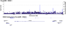
We probably discussed this already but in DecodeME there wasn't a strong signal around TPRM3 (although a weak one at 9:71403764. P Value: 2.29 x 10^-3).

Thanks for reminding us. If there was an interesting contribution from TRMP3 genetics I would expect DecodeME to have come up with something crisper. I may be wrong, but it looks as if the signal originally claimed for TMP3 is not replicating.
 
There was some discussion in the welcome thread for James Cox of looking at these findings again (I think specifically the TRPM3 part) to see if there was anything real there.

I know @hotblack has been looking at NK cells having some sort of 'memory' lately (havent read the paper yet so am probably getting it wrong) - could that tie in at all?

I know there's a lot of skepticism around this teams findings and I share it, just wondering if we're missing anything.
 
We probably discussed this already but in DecodeME there wasn't a strong signal around TPRM3 (although a weak one at 9:71403764. P Value: 2.29 x 10^-3).
Also, here is the most significant SNP from the paper which I think started their interest specifically in TRPM3, and the p-value from that paper (Marshall-Gradisnik 2015): rs12682832, p=0.003

Looking up this SNP in DecodeME, it's about as non-significant as could be (p=.97):
1779725849638.png

So it seems likely that the signal in that genetic study was a false positive, caused by lack of multiple test correction.

---

As for the ongoing TRPM3 lab studies, it seems important to figure out what is happening. Either it's a real, and apparently very reliable effect, in which case it seems important that it gets followed up on, or their lab has been doing something wrong in many studies for years, in which case it's important to know so that resources don't get wasted on this going forward.
 
As for the ongoing TRPM3 lab studies, it seems important to figure out what is happening. Either it's a real, and apparently very reliable effect, in which case it seems important that it gets followed up on, or their lab has been doing something wrong in many studies for years, in which case it's important to know so that resources don't get wasted on this going forward.
Agreed, we need a trusted team to attempt replication.

It would be good if we could get someone like Audrey Ryback to do a thorough job of it.

We need to either follow this where it leads or put it to bed for good.
 
I've never used GraphPad, so I don't know what exactly "GraphPad formed statistical comparison by participant, not by individual cell" means. If it means the values from each participant were averaged, that would be sufficient to avoid pseudoreplication, but it would be preferable to see more details about what exactly was done.
I’ve only used GraphPad sparingly but looking at some of the violin plots, getting p<0.0001 with a very small median difference and near complete overlap using Mann-Whitney U-test on N=10 seems unusual, to put it lightly. Unless data was explicitly averaged by participant I have no idea how pseudoreplication could have been avoided here with that specific test.

Presumably the calcium flux in the NK cells in people with ME/CFS isn't a clinical problem. So at best it might be a clue to a calcium flux problem somewhere more interesting. But again, we don't seem to have much evidence for that.
That’s my sense too. It may speak to NK cells being transiently exposed to some signaling and TRPM3 is one of several proteins affected.

Though I’ve done a re-analysis of the published Vu et al. scRNA-seq PBMC data (will be doing a poster at Invest in ME soon) and did not see much of a strong transcriptional difference overall in NK cells, definitely no strong signals in any calcium signaling-related pathways. If it’s real it seems like a minor functional difference that only becomes apparent with the experimental stimulation here.
 
Back
Top Bottom