List of causation hypothesis for follow up after DecodeME

Kiristar

Senior Member (Voting Rights)
Coming back up out of the detail we could really do with leveraging the Decode ME and other recent findings and working out a research roadmap which we could perhaps refine at the showcase event to develop the what next plan?

Someone on the initial findings thread suggested developing a list of hypotheses, which feels to me like the next step towards that.

So I thought I would create a new thread so any hypothesis ideas don't get lost in that very detailed thread.

Hope that is a workable approach and makes sense to people ....?
 
Last edited by a moderator:
I think it would be worthwhile to talk about the hypotheses or areas of investigation we find the most compelling. An offical S4ME list could perhaps be contentious and require a vote to make it fair, but at least in an informal fashion i think it would be a good idea.
 
Last edited:
My original post in the main DecodeME thread that was referenced in Kiristar's post above, for reference.

Another thought: Would it be worthwhile to create a list of the hypotheses we think are worth pursuing, both by members here and by others? In a members only post or even a private group. We could then speculate on what experiments could be done to validate/falsify these hypotheses.
 
My view on this is that there are some broad hypothesis options worth highlighting in the wake of DecodeME and some maybe to put aside.

1. The potential significance of a gamma delta T cell related gene strengthens the case for more functional T cell studies along the lines that Jackie Cliff has been exploring. Forgetting the FcRI idea from our Qeios paper I think there is a case for 'a little more T cells'. The hypothesis would be broadly that by analogy with psoriasis or Reiter's syndrome ME/CFS is at least in part a persistent T cell activation problem, maybe most likely involving rather 'innate' populations like MAIT or gamma delta. They would need to some sort of 'covert' population in terms of their trafficking and effector role. (The paper posted here on MAIT cells going to meninges is intriguing.) The practical idea would be to replicate the sort of work Jackie has been doing with a focus on looking for priming of circulating T cells. Looking for T cells in the head might be justified but I doubt it would be easy.

2. The CA10 locus, apparently shared with chronic pain suggests that there may be a susceptibility to an amplification loop involving synapses that is relevant both to pain and other ME/CFS symptoms. How you investigate that beyond more refined genetics I am not sure. I just wonder whether it would be worth looking for links with migraine on the basis that that seems to be another situation where a brain loop generates signals that should not be there.

3. Where 1. focuses on T cell overactivity and maybe interferon gamma production I think the idea that interferon alpha acting locally might be more important long term, perhaps from dendritic cells or from 'trained' tissue macropahges or microglia is worthy of attention. As jnmaciuch has pointed out, this might make sense of several DecodeME hits. Microglial activation studies make sense, even if I am pessimistic that they will show much with current methods.

Areas DecodeME would encourage me further to move away from include autoantibodies, mitochondrial energy supply and persistent viruses. (Jackie's focus does involve persistent herpes viruses but I think the significance of her studies may go beyond any specific relation to herpes.) But there's no surprises there.
 
The research and hypotheses I am most interested in currently:

The Edwards/Cambridge/Cliff Hypothesis, if it still stands post DecodeME, and whatever emerges like a phoenix from its ashes if it doesn't.

@jnmaciuch's interest in type 1 interferons, and interferon as a possible lead generally.

Fluge and Mella's ongoing work on Daratumumab and llpc depletion.

Paul Hwang's work on wasf3.

The recent 3d skeletal muscle paper by Mughal et al.
 
Last edited:
If there is persistent T cell activation would we expect to see more gramzyme B and perforin in the blood? I think from studies certain T cell types don't make as much of the two when tested in the lab.

According to the Selin lab only a subset show gamma-delta T cell differences. I suspect identifying subsets and testing longitudinally will be important in researching T cells. There is also the debate of fresh vs frozen cells and sorting methods and how they affect results. It's not as easy as it sounds to do a deep dive with large sample numbers. I'm sure what Dr. Cliff learns about longitudinal sampling will be helpful for others moving forward.

Anyone know if Dr Selin and Dr Cliff meet up to brainstorm T cells? Might be worthwhile given the DecodeME pointers.

From my experience of being a research subject it takes a lot of tubes of blood to have enough of some of the rarer type T cells......
 
The paper posted here on MAIT cells going to meninges is intriguing.
It has nothing to do with trafficking, unfortunately. MAIT were found in the meninges in healthy conditions, as were other T cell subsets. It was the lack of MAIT that lead to increased meningeal barrier permeability, which they said was due to a lack of MAIT-secreted free radical scavengers (where ROS seemed to interfere with cell-cell junctions in the barrier). And they showed nothing about T cells crossing the barrier themselves.
 
But why only hypotheses? For a Roadmap couldn't there be also be a host of sensible genetic work or follow-up work that can also be entirely hypothesis free?
I was just thinking that projects test hypotheses so was thinking you'd start there then move to designing projects as an outcome of knowing what we need to answer next. But if a different method to get to the projects works I'm all for it.
 
I am intending to look along the lines of how specific mechanisms that may lead to altered ER-mitochondria interactions (including by biogenesis or phagy of either, lipid content of or lipid shuttling by either, calcium storage or mobilisation, etc) may interact with activation of or signalling by particular immune cell subsets. But this depends on funding. I have samples, collaborators, techniques and facilities in place to do all of this but need $ to do it. Writing proposals.

PS i already have a funded study (halfway in) that’s looking at T cell activation in collaboration with T cell experts. stay tuned
 
Last edited:
@DMissa not sure if this is the right place to post this, but if you’re doing any high throughput screeen automation I’m happy to donate my time. I’m an automation engineer with 7+ years of experience in biotech specializing in high throughput screening on liquid handlers: Hamilton, Beckman (Bravos & echo), Tecans, thermo-fisher work cells, and custom integration work cells.

I’ve ask OMF in the past but have learned it’s cheaper outside of biotech to just hire grad students to pipette instead of buying robotics. If anyone working on CFS needs automation consulting happy to donate my time.
 
The following hypothesis was generated using a multi-reasoning approach which uses 5 #AI reasoning engines.

multi-reasoning.png

The latest hypothesis is as follows given the input data. Note that the model includes ways to check the validity of this hypothesis :

- ME/CFS is a genetically primed failure to extinguish an interferon-coupled integrated stress response after a trigger (infection or non-viral/toxic). Mitochondrial and ER damage release danger nucleic acids and misfolded proteins that activate RLR/cGAS–STING and type I/III interferons. Defects in vesicle trafficking and ER‑phagy prevent proper localization and shutdown of these pathways and of IFN receptor signaling. The result is a chronic, low‑grade IFN tone that sustains the ISR (driving elevated FGF21), shifts lipid metabolism toward phospholipid peroxidation and depletion, and propagates immune, microvascular, and neuro-autonomic dysfunction.

Why this fits the genetics (priority-coded)
- GWAS (highest priority)
- RABGAP1L, ARFGEF2: Endosome–Golgi–plasma membrane trafficking and receptor recycling. Control STING/TLR movement and IFNAR endocytosis/recycling; impaired trafficking → prolonged or mislocalized IFN signaling and poor resolution.
- CCPG1: ER‑phagy receptor clearing stressed ER membranes that harbor STING/TLR complexes; inefficiency → sustained IFN and UPR activity.
- FBXL4: Mitochondrial DNA/biogenesis maintenance; distress releases mtDNA/RNA that drive cGAS–STING/RLR and upregulates mitokines like FGF21.
- SUDS3: SIN3/HDAC corepressor component shaping global stress/immune transcription, including ISGs; variants bias cells toward persistent “conservation” programs.
- BTN2A2: Butyrophilin co‑regulator of T and γδ T cells, tuning IFN‑γ and co‑stimulation set‑points.
- OLFM4: Neutrophil/gut crypt factor linking mucosal innate immunity and type III IFN (IFN‑λ) at barrier sites.
- CA10: Brain-enriched synaptic modulator; explains central/autonomic phenotypes in the setting of systemic IFN/ISR and vascular change.
- Tier 1 (high-confidence colocalized; highlights by pathway)
- IFN sensors/regulators: ZNFX1 (dsRNA sensing/mtRNA handling), TRIM38 (E3 ligase gating TLR/cGAS–STING amplitude/termination), RC3H1/Roquin (post‑transcriptional braking of immune mRNAs), ZBTB37/ZNF322/HMGN4 (chromatin states of ISGs).
- Trafficking/clearance: KLHL20 (Cullin3 E3; autophagy programs), CSE1L (nuclear/secretory transport), B4GALT5 (glycosphingolipid rafts affecting TLR/IFN signaling domains), STAU1 (stress granules that scaffold RLR signaling), DDX27/ABT1 (ribosome biogenesis coupling to antiviral translation control), DDX27 also impacts nucleolar stress.
- Mitochondrial/ISR: DARS2 (mt-aaRS; mitochondrial translation stress → ISR/FGF21), ZNFX1 as above.
- Lipid redox and eicosanoids: PRDX6 (peroxidase/iPLA2; repairs yet consumes phospholipids), PEBP1/RKIP (enables 15‑LOX–PE peroxidation; also modulates NF‑κB/MAPK), HFE (iron redox), PTGIS (prostacyclin synthase; endothelial tone), SERPINC1 (antithrombin; thromboinflammation).
- Immune co-stimulation/barrier: TNFSF4/OX40L, BTN3A3, VSIG10; SLC9C2 (endosomal/epithelial pH influencing TLR compartments).
- Additional: GPR52 (CNS cAMP; autonomic modulation), PEBP1 and PEBP1–15‑LOX ferroptotic signaling interface with lipid peroxidation.
- Tier 2 (nearest-gene where Tier 1 was absent): FBXL4, OLFM4, CCPG1 (also GWAS hits) reinforce the mitochondrial, mucosal, and ER‑phagy axes.

Mechanistic chain (now explicit for IFN)
1) Trigger (virus, bacterial products, organophosphates/toxins, ischemic stress) perturbs membranes, Ca2+, and redox → ER misfolding and mitochondrial ROS/damage.
2) Mito‑nucleic acid release activates RLRs and cGAS–STING → type I IFN (α/β); mucosal sites emphasize type III IFN (λ). T/NK circuits contribute type II IFN (γ) depending on BTN/OX40L set‑points.
3) Trafficking and ER‑phagy resolution fails: ARFGEF2/RABGAP1L mis-handle STING/TLR/IFNAR trafficking; CCPG1‑dependent ER‑phagy clears poorly. TRIM38/RC3H1/ZNFX1/ STAU1/DDX27 set abnormal on/off kinetics. Net effect: tonic, spatially mislocalized IFN signaling.
4) IFN–ISR coupling: dsRNA‑PKR and PERK phosphorylate eIF2α; translation throttles, ATF4/CHOP programs engage, and FGF21 rises as a systemic mitokine.
5) Lipid injury and depletion: PRDX6 and PEBP1–15‑LOX drive phospholipid peroxidation and lyso‑PL generation; HFE‑linked iron accelerates lipid ROS. B4GALT5‑dependent raft changes alter TLR/IFN microdomains. Plasma phosphatidylcholines/PE fall, oxidized/lyso‑PLs rise.
6) Vascular/coagulation: PTGIS and SERPINC1 axes shift toward endothelial dysfunction, platelet activation, and microclots, impairing oxygen delivery and recovery after exertion.
7) Immune set‑point: BTN2A2/BTN3A3 and TNFSF4/OX40L reshape T and γδ T‑cell circuits in a background of tonic IFN, favoring poorly resolving inflammation and post‑exertional exacerbations.
8) CNS/autonomic: CA10/GPR52 plus systemic IFN/ISR and microvascular change yield cognitive, sensory, and orthostatic symptoms.

Why non‑viral triggers fit
- Toxins such as organophosphates can cause mitochondrial/ER damage and lipid peroxidation, releasing DAMPs (mtDNA/RNA) that drive cGAS–STING/RLR and IFN without infection. The shared bottleneck is resolution capacity (trafficking + ER‑phagy), not the trigger’s origin.

Key predictions (falsifiable)
- Whole blood or single-cell ISG signatures show tonic elevation with altered kinetics (prolonged TBK1/IRF3–STAT1/2 signaling, blunted receptor recycling).
- Patient cells display delayed STING trafficking to lysosomes and impaired shutdown; defects correlate with ARFGEF2/RABGAP1L alleles; CCPG1 loss-of-function phenotypes show reduced ER‑phagy flux.
- Post‑exertion, transient spikes in cell‑free mtDNA and type I/III IFNs accompany rises in FGF21 and lipid peroxidation adducts (e.g., 4‑HNE; 15‑LOX–PE species).
- Plasma lipidomics: reduced PCs/PEs, elevated lyso‑PLs/oxidized‑PLs; altered glycosphingolipids (B4GALT5).
- Endothelial tests: lower prostacyclin metabolites, antithrombin imbalance, increased thromboinflammatory/microclot markers.
- Gut-dominant phenotypes show stronger IFN‑λ signatures and OLFM4‑linked neutrophil/NET activity.

Implications (research directions, not clinical advice)
- Restore resolution of IFN signaling: target STING/TLR trafficking and degradation (autophagy/lysosomal enhancers; carefully titrated STING modulators); bolster CCPG1‑axis ER‑phagy.
- Tune the ISR rather than blanket immunosuppression: timed ISR modulators; support mitochondrial translation/biogenesis in genetically susceptible backgrounds (e.g., FBXL4/DARS2 contexts).
- Repair membrane composition and limit ferroptotic chemistry: plasmalogen/phospholipid repletion, inhibitors of PEBP1–15‑LOX complexes, iron handling support; antioxidant strategies targeted to membranes.
- Improve microvascular tone/antithrombotic balance: prostacyclin pathway, antithrombin support, careful platelet modulation.
- Calibrate costimulatory axes: BTN/OX40L pathway research to reset T/γδ T‑cell set‑points without ablating host defense.

In short: ME/CFS may be a “failure to extinguish IFN‑coupled stress” disorder. Genetic variation in trafficking (RABGAP1L, ARFGEF2), ER‑phagy (CCPG1), mitochondrial maintenance/translation (FBXL4, DARS2), transcriptional control (SUDS3), and immune set‑points (BTN2A2 and others) biases recovery toward a chronic state with tonic IFN, sustained ISR (FGF21‑high), and phospholipid injury (phospholipids‑low), producing the multi‑system phenotype.

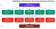
and the proposed interventions (which have many commonalities with the regimen I originally used. Note the mentions on lipid peroxidation, ER Stress and the combination of ER Stress control, Oxidative stress and cell membrane support :

Combination and sequencing (pragmatic)

  • Phase 1 (4–6 weeks): Foundations + lipid-peroxidation block and membrane repletion (vitamin E/C + selenium + CoQ10 + PC/citicoline ± NAC/ALA) + MCAS control if present.
  • Phase 2 (6–8 weeks): Add an ER-stress modulator (TUDCA or 4-PBA) + LDN. Titrate slowly; track FGF21 and symptom fluctuation.
  • Phase 3: Address dominant phenotype
    • Orthostatic/microvascular: add pentoxifylline ± sulodexide; optimize OI meds; consider low-dose aspirin.
    • Sticky innate/T cell: trial hydroxychloroquine or colchicine microdose (not together initially).
    • Energy ceiling: add carnitine/creatine/D-ribose; consider ketone support if GI allows.
  • Phase 4 (specialist/clinical trial tier): sirolimus microdosing, prostacyclin-pathway agents, IVIG for defined immune deficits, antivirals only with evidence of active replication, elamipretide or deuterated PUFAs via studies.
What to avoid or minimize (fits the model)

  • High-heat/reused oils and very high PUFA loads without antioxidants; severe caloric restriction/extended fasting; repeated “crash” exertion; organophosphate/carbonate pesticide exposure; unnecessary immune activators (e.g., chronic stimulant use if it worsens PEM).
Biomarker-guided care

  • Core: FGF21 (ISR load), CRP and a limited cytokine panel, lipid peroxidation marker (F2-isoprostane), fasting lipidomics if accessible, orthostatic testing, actigraphy/HR variability.
  • Optional: RBC deformability, platelet activation/microclot staining (if standardized), ISG score, ATF4 target panel from PBMCs.
Safety notes

  • Start low, go slow; introduce one new agent every 2–4 weeks with a clear stop rule.
  • Watch for drug–drug interactions (e.g., SSRIs with other serotonergic agents; anticoagulants/antiplatelets with NSAIDs).
  • Monitor liver enzymes with TUDCA/4-PBA; renal function with metformin-type ideas; eye exams with hydroxychloroquine; bleeding risk with vascular agents.
If you can share which features dominate (e.g., FGF21 levels, lipidomics, presence of OI/microvascular signs, GI/MCAS symptoms), I can help prioritize a tighter, step-by-step plan and pick specific starting combinations.
 
I believe the reason LLMs are useless here is because they repeat the speculations of others with no ability to discern sense from nonsense, and mix in knowledge of how things are connected in the body as well, with little ability to figure out what's actually relevant. If among these speculations were ideas that generally worked well, one would expect the accumulation of evidence supporting them.
 
It has nothing to do with trafficking

I don't follow that. Presumably the MAIT cells did traffic there - which was the point. The apparently beneficial effect is peculiar but of course if these cells are having some sort of negative effect that could be just as relevant. I don't see reactive oxygen species as being of particular interest to pathology in ME/CFS. I am not even sure that meninges are very relevant to ME/CFS but if MAIT cells can get there they might have an influence on brain function
 
If there is persistent T cell activation would we expect to see more gramzyme B and perforin in the blood?

A little while back I posted a query about circulating T cell findings in things like psoriasis. I am not sure one expects to find much. There need be no obvious shifts in population numbers. In fact the bad cells are likely to be absent from circulation, doing bad things elsewhere.

My thought was that you might pick something up by looking at how circulating T cells interact with other cells in short term culture. That may still not get over the problem but if people don't find much in diseases like psoriasis where we know there are misbehaving T cells then finding nothing in ME/CFS does not rule a T cell-mediated problem.
 
I was looking up pathogenesis of Parkinson's Disease and asked Google and also PubMed. The result is instructive. Google mentions two risk genes but presents them in a way that makes no real sense. That is presumably because review articles have given plain English summaries of the roles for these genes that do not actually reflect the complex dynamics usefully. So Google tries to be clever and compare them and comes up with something that makes no sense.

The reviews are interesting in that they focus on specific pathological findings, like accumulation of alpha synuclein in granules in dopaminergic cells. But there is very little discussion of the systems dynamics that would fit the findings into a story to explain the genetics, the age range, the rate of onset and so on. The structure of the systems dynamics seems to me to be an essential starting point for building a plausible model. That was very much what Robert Phair was arguing for his shunt and we have argued over subsequently with interferons. What sort of process would manifest as spreading appearance of misfolded protein aggregates in. particular cell type. To me it seems very much like a prion story, but linked to some intrinsic system failure that only ever occurs after several decades (there are some very rare exceptions). I suspect that the environment is a red herring.

Having a workable model of dynamic structure may not necessarily affect your choice of treatment. We got the dynamics of RA a bit wrong on plasma cell longevity initially but rituximab still worked. But I think for ME/CFS having at least a high level model of overall dynamic would make it much easier to justify candidate treatments.

The problem with writing papers about system dynamics of disease is that, by and large, referees don't understand and reject the paper. So I can see why there are so few reviews that go into it.
 
Talked about an idea in this thread - Could nerve damage or retrograde microtubule based transport in axons explain the delay associated with PEM? about whether the time delay associated with PEM could be linked to the amount of time it takes from nerve injury/an event at nerve terminals to the nucleus generating a transcriptional response. This was based off the possibility that organophosphates, which cause certain neurons to fire constantly and would damage synapses, seem to be able to trigger an ME like illness.

I've learnt since then that apparently some forms of synaptic plasticity - namely synaptic scaling - also occur on timescales that line up well with PEM (hours to days). This is a homeostatic mechanism to keep global firing rates of a neuron relatively stable, such that if a neuron has been firing a lot due to stimulation at any of its synapses it will begin to scale down - and vice versa (The Self-Tuning Neuron: Synaptic Scaling of Excitatory Synapses, Turrigiano 2010)

I think it's also worth considering protein degradation, which has come up in a few forms now. The proteasome in Zhang HEAL2, and autophagy is plausibly linked to at least three genes in decode (FBXL4, CCPG1, RABGAP1L). Protein degradation appears to be a key part of synapse homeostasis.

2. The CA10 locus, apparently shared with chronic pain suggests that there may be a susceptibility to an amplification loop involving synapses that is relevant both to pain and other ME/CFS symptoms. How you investigate that beyond more refined genetics I am not sure. I just wonder whether it would be worth looking for links with migraine on the basis that that seems to be another situation where a brain loop generates signals that should not be there.

In my opinion there are a bunch of things that point to synapses, but how clear is it really that there would be amplification, rather than attenuation or something else. It does seem like it could be quite complicated - if a lot of a neuron's synapses at the dendrites were compromised for example and connections were lost, maybe the neuron could end up firing more (erratically) later anyway due to a mechanism such as synaptic scaling as above.
 
In my opinion there are a bunch of things that point to synapses, but how clear is it really that there would be amplification, rather than attenuation or something else. It does seem like it could be quite complicated - if a lot of a neuron's synapses at the dendrites were compromised for example and connections were lost, maybe the neuron could end up firing more (erratically) later anyway due to a mechanism such as synaptic scaling as above.

I can definitely buy that.
Amplification was probably not the best word. Something is wrong with control mechanisms but as you say, it could be quite complicated!
 
Last edited:
Talked about an idea in this thread - Could nerve damage or retrograde microtubule based transport in axons explain the delay associated with PEM? about whether the time delay associated with PEM could be linked to the amount of time it takes from nerve injury/an event at nerve terminals to the nucleus generating a transcriptional response. This was based off the possibility that organophosphates, which cause certain neurons to fire constantly and would damage synapses, seem to be able to trigger an ME like illness.

I've learnt since then that apparently some forms of synaptic plasticity - namely synaptic scaling - also occur on timescales that line up well with PEM (hours to days). This is a homeostatic mechanism to keep global firing rates of a neuron relatively stable, such that if a neuron has been firing a lot due to stimulation at any of its synapses it will begin to scale down - and vice versa (The Self-Tuning Neuron: Synaptic Scaling of Excitatory Synapses, Turrigiano 2010)

I think it's also worth considering protein degradation, which has come up in a few forms now. The proteasome in Zhang HEAL2, and autophagy is plausibly linked to at least three genes in decode (FBXL4, CCPG1, RABGAP1L). Protein degradation appears to be a key part of synapse homeostasis.


In my opinion there are a bunch of things that point to synapses, but how clear is it really that there would be amplification, rather than attenuation or something else. It does seem like it could be quite complicated - if a lot of a neuron's synapses at the dendrites were compromised for example and connections were lost, maybe the neuron could end up firing more (erratically) later anyway due to a mechanism such as synaptic scaling as above.
I’m not a neuroscience person so maybe I’m missing some important context—but if it had to do with something traveling down a neuron that wasn’t an action potential, wouldn’t you expect the progression of PEM to be more, well, progressive across the body based on shortest to longest axonal length? I definitely get whole-body muscle weakness, stiffness, and pain as part of PEM and sometimes symptoms have a temporal offset between them, but not in any way that would correspond to neuron length. For me, it’s more that pain/stiffness/weakness is the worst in the muscle groups that I was using during activity, and then everywhere else it builds at the same rate and around the same intensity.
 
Back
Top Bottom