Stress-inducible WASF3 regulates brain metabolism and cognitive function 2026 Syed, Hwang et al

Andy

Senior Member (Voting rights)
Author list: Abu M. Syed, Jin Ma, Ping-yuan Wang, Youlim Son, Rozhin Penjweini, Nikhil Mattu, Jay R. Knutson, Sachiko Nakamori-Yamashita, Danielle A. Springer, Ananya Ganguly, Chengyu Liu, and Paul M. Hwang

Significance​

Cognitive impairment, often described as “brain fog,” is a disabling feature of chronic fatigue syndrome (ME/CFS), but its underlying mechanism remains unclear. This study shows that stress-inducible Wiskott–Aldrich syndrome protein family member 3 (WASF3), previously linked to mitochondrial dysfunction in skeletal muscle of patients with ME/CFS, also suppresses oxidative metabolism in the brain. Increased WASF3 shifted hippocampal metabolism toward a hypometabolic state and impaired learning. Conversely, reduced WASF3 enhanced oxidative metabolic markers and protected cognitive function during cellular stress. These findings identify WASF3 as a mediator linking endoplasmic reticulum (ER) stress to brain bioenergetic dysfunction and suggest that targeting ER stress-WASF3 signaling may alleviate cognitive symptoms in ME/CFS and related postviral fatigue disorders more broadly.

Abstract​

Wiskott–Aldrich syndrome protein family member 3 (WASF3) disrupts mitochondrial respiration and is induced in association with endoplasmic reticulum stress in myalgic encephalomyelitis/chronic fatigue syndrome (ME/CFS). Because cognitive impairment is a hallmark of ME/CFS, we investigated the role of WASF3 in brain metabolism and function using WASF3 transgenic and knockout mouse models. WASF3 disrupted respiratory complex IV and localized to mitochondria-associated membranes, known to modulate cellular processes important for neuronal activity. Overexpression of WASF3 in the hippocampus, a critical region for memory formation, induced a metabolic shift toward glycolysis and promoted a “hypometabolic” state. Increased WASF3 also caused a reduction in ATPIF1, previously shown to facilitate cognitive function, and impaired learning. Conversely, WASF3 deficiency increased ATPIF1 level, enhanced oxidative metabolism, and preserved cognitive function under stress. Together, these findings demonstrate that WASF3 suppresses oxidative metabolism in the brain and contributes to cognitive dysfunction, providing mechanistic insights into the “brain fog” reported in ME/CFS.

Open access
 
Going only by the abstract, their hypothesis is that of increased WASF3 in pwMECFS? Which should mean we should see more glycolysis and less oxidative metabolism? But haven't we previously seen the opposite, i.e. less glycolysis and more oxidative metabolism? Or is this only brain specific, and doesn't address what might be going on elsewhere?

Edit to add: Yes, I appreciate this study looks at the brain only. I suppose my question is, is it possible to have less glycolysis in the rest of the body, and increased in the brain, in order for both findings to be possible?
 
Last edited:
Andy, would this address the question about the involvement of glycolysis vs oxidative metabolism in ME/CFS?

"Studies have found increased glycolytic activity and reduced efficiency of mitochondrial oxidative phosphorylation in CFS patients, leading to energy deficiency and the accumulation of metabolic waste, such as lactate."

"PKM2 plays a crucial role in this process. PKM2 is a key enzyme in the glycolytic pathway, catalyzing the conversion of phosphoenolpyruvate to pyruvate, generating ATP."

"Research suggests that abnormal activation of PKM2 in hippocampal cells may enhance glycolysis and lactate accumulation, leading to mitochondrial dysfunction and accelerating the progression of CFS. Furthermore, the activation of PKM2 not only promotes glycolysis but also regulates lactate accumulation, which further alters the acidic environment within cells. This metabolic disorder is considered a crucial mechanism underlying energy production deficits in CFS patients."

 
Yes, I appreciate this study looks at the brain only. I suppose my question is, is it possible to have less glycolysis in the rest of the body, and increased in the brain, in order for both findings to be possible?
Yes the metabolism in one cell type doesn’t necessarily tell you anything about another. Though I think any findings of a shift in glycolysis/oxidative metabolism in tissues that have been studied in ME/CFS were largely inconsistent between themselves to begin with
 
Andy, would this address the question about the involvement of glycolysis vs oxidative metabolism in ME/CFS?

"Studies have found increased glycolytic activity and reduced efficiency of mitochondrial oxidative phosphorylation in CFS patients, leading to energy deficiency and the accumulation of metabolic waste, such as lactate."

"PKM2 plays a crucial role in this process. PKM2 is a key enzyme in the glycolytic pathway, catalyzing the conversion of phosphoenolpyruvate to pyruvate, generating ATP."

"Research suggests that abnormal activation of PKM2 in hippocampal cells may enhance glycolysis and lactate accumulation, leading to mitochondrial dysfunction and accelerating the progression of CFS. Furthermore, the activation of PKM2 not only promotes glycolysis but also regulates lactate accumulation, which further alters the acidic environment within cells. This metabolic disorder is considered a crucial mechanism underlying energy production deficits in CFS patients."

Thanks, we have a thread on that here.
 
Overexpression of WASF3 in the hippocampus, a critical region for memory formation, induced a metabolic shift toward glycolysis and promoted a “hypometabolic” state. Increased WASF3 also caused a reduction in ATPIF1, previously shown to facilitate cognitive function, and impaired learning. Conversely, WASF3 deficiency increased ATPIF1 level, enhanced oxidative metabolism, and preserved cognitive function under stress. Together, these findings demonstrate that WASF3 suppresses oxidative metabolism in the brain and contributes to cognitive dysfunction, providing mechanistic insights into the “brain fog” reported in ME/CFS.
Did WASF3 show up in DecodeME analysis? I don't remember it.
 
Did WASF3 show up in DecodeME analysis? I don't remember it.
I don't think it did, but maybe the association is between a regulatory sequence to WASF3 and ME/CFS, rather than WASF3 itself and ME/CFS.

A regulatory sequence affects how much of the gene product is made. That could lead to a tendency to overexpress WASF3 and be more likely to suffer the disruption of energy metabolism.

WASF3 could also be dysregulated because the proteins that regulate it are dysregulated.
 
Last edited:
To assess the effect of WASF3 overexpression on cognitive function, we used fear conditioning, a widely employed paradigm of associative learning that is quantified by motionless (freezing) time upon re-exposure to a context or discrete cue (e.g., a tone) in association with an aversive stimulus. WASF3 Tg mice showed decreased freezing time, particularly with the cued fear conditioning paradigm. Because specific types of contextual and cued fear conditioning depend on different parts of the brain, these data indicated that overexpression of WASF3 in the brain could compromise behavior by affecting mitochondrial metabolism and ΔΨ m that underlie neuronal signaling.

These findings support a model in which dysregulated WASF3 transduces cellular stress signals into mitochondrial dysfunction, contributing to the cognitive alterations observed in ME/CFS. Our observation of the WASF3-mediated changes in fear conditioning behavior, a key component of the posttraumatic stress disorder (PTSD) associated with ME/CFS (40–42), further underscored ER stress-WASF3 signaling as a potential contributing mechanism in this disorder.

[40] is The Comorbidity of Self-Reported Chronic Fatigue Syndrome, Post-Traumatic Stress Disorder, and Traumatic Symptoms (2012, Psychosomatics)
BACKGROUND
Data from primary care and community samples suggest higher rates of post-traumatic stress disorder (PTSD) among individuals with chronic fatigue syndrome (CFS).

OBJECTIVE
This study investigated the co-occurrence of CFS, PTSD, and trauma symptoms and assessed the contribution of familial factors to the association of CFS with lifetime PTSD and current traumatic symptoms.

METHOD
Data on lifetime CFS and PTSD, as measured by self-report of a doctor's diagnosis of the disorder, and standardized questionnaire data on traumatic symptoms, using the Impact of Events Scale (IES), were obtained from 8544 female and male twins from the community-based University of Washington Twin Registry.

RESULTS
Lifetime prevalence of CFS was 2% and lifetime prevalence of PTSD was 4%. Participants who reported a history of PTSD were over eight times more likely to report a history of CFS. Participants with scores ≥ 26 on the IES were over four times more likely to report CFS than those who had scores ≤ 25. These associations were attenuated but remained significant after adjusting for familial factors through within-twin pair analyses.

CONCLUSION
These results support similar findings that a lifetime diagnosis of CFS is strongly associated with both lifetime PTSD and current traumatic symptoms, although familial factors, such as shared genetic and environmental contributions, played a limited role in the relationship between CFS, PTSD, and traumatic symptoms. These findings suggest that future research should investigate both the familial and the unique environmental factors that may give rise to both CFS and PTSD.

[41] is Understanding posttraumatic stress disorder through fear conditioning, extinction and reconsolidation (2016, Neuroscience & Biobehavioral Reviews)
Posttraumatic stress disorder (PTSD) is a psychopathology characterized by exacerbation of fear response. A dysregulated fear response may be explained by dysfunctional learning and memory, a hypothesis that was proposed decades ago.

A key component of PTSD is fear conditioning and the study of this phenomenon in laboratory has expanded the understanding of the underlying neurobiological changes in PTSD. Furthermore, traumatic memories are strongly present even years after the trauma and maintenance of this memory is usually related to behavioral and physiological maladaptive responses.

Persistence of traumatic memory may be explained by a dysregulation of two memory processes: extinction and reconsolidation. The former may explain the over-expression of fear responses as an imbalance between traumatic and extinction memory. The latter, in turn, explains the maintenance of fear responses as a result of enhancing trauma-related memories.

Thus, this review will discuss the importance of fear conditioning for the establishment of PTSD and how failure in extinction or abnormal reconsolidation may contribute to the maintenance of fear response overtime.

HIGHLIGHTS
• Fear conditioning plays a major role in PTSD.

• The main areas involved in fear conditioning are also altered in PTSD.

• Persistence of traumatic memory could be a failure of extinction learning.

• Exacerbated fear may also be a consequence of enhanced trauma-related memories through reconsolidation.

[42] is Post-Traumatic Stress Disorder and Complex Post-Traumatic Stress Disorder in people with long COVID, ME/CFS, and controls (2023, The American Journal of Medicine)
BACKGROUND
Prevalences of Post-Traumatic Stress Disorder (PTSD) and Complex Post-Traumatic Stress Disorder (CPTSD) have not previously been compared between individuals with long COVID and individuals with Myalgic Encephalomyelitis/ Chronic Fatigue Syndrome (ME/CFS), and healthy age-matched controls. For these reasons, this study aimed to determine the prevalence of PTSD and CPTSD in individuals with long COVID (n=21) and ME/CFS (n=20) and age-matched controls (n=20).

METHODS
A case-case-control approach was employed, participants completed the International Trauma Questionnaire (ITQ), a self-report measure of the International Classification of Diseases (ICD-11) of PTSD and CPTSD consisting of 18 items. Scores were calculated for each PTSD and Disturbances in Self-Organization (DSO) symptom cluster and summed to produce PTSD and DSO scores. PTSD was diagnosed if the criteria for PTSD were met but not DSO, and CPTSD was diagnosed if the criteria for PTSD and DSO were met. Moreover, each cluster of PTSD and DSO were compared among individuals with long COVID, ME/CFS and healthy controls.

RESULTS
Individuals with long COVID (PTSD= 5%, CPTSD=33%) had more prevalence of PTSD and CPTSD than individuals with ME/CFS (PTSD= 0%, CPTSD=20%) and healthy controls (PTSD= 0%, CPTSD=0%). PTSD and CPTSD prevalence was greater in individuals with long COVID and ME/CFS than controls. Individuals with long COVID had greater values controls for all PTSD values. Moreover, individuals with long COVID had greater values than controls for all DSO values. Individuals with ME/CFS had greater values than controls for all DSO values. Both long COVID and ME/CFS groups differed in overall symptom scores compared to controls.

CONCLUSIONS
Findings of this study demonstrated that individuals with long COVID generally had more cases of PTSD and CPTSD than individuals with ME/CFS and healthy controls.
 
The PTSD profile on chromatin opening caught my attention in one of the recently published comparative tables. It mostly refer to non-neuronal cells, although ME/cfs refer to neurons.
Link
So, I’m wary of comparisons with TSPT and ME/cfs. Perhaps the only common link is sleep disorders.

--Incidentally, the table is rather amusing: some so-called neurological disorders involve in fact non-neuronal cells, whilst some so-called ‘psychiatric’ disorders involve neurons.

The problem is that non-neuronal cells can include, amongst others, astrocytes and glial cells, and PTSD could in fact be a condition with real inflammation; I feel that the trauma-based approach is, to say the least, open to question.
 
Last edited:
Going only by the abstract, their hypothesis is that of increased WASF3 in pwMECFS? Which should mean we should see more glycolysis and less oxidative metabolism? But haven't we previously seen the opposite, i.e. less glycolysis and more oxidative metabolism? Or is this only brain specific, and doesn't address what might be going on elsewhere?

Edit to add: Yes, I appreciate this study looks at the brain only. I suppose my question is, is it possible to have less glycolysis in the rest of the body, and increased in the brain, in order for both findings to be possible?
Andy, I can't find any studies that say that ME/CFS involves more oxidative metabolism and less glycolysis. Where did you see that?
Thank you.
 
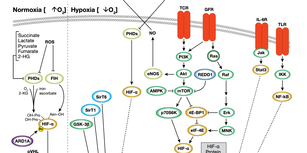
Maybe hypoxia is the upstream event.

"In particular, WASF3 is a hypoxia-inducible gene,"

"meaning that it contains at least one or more hypoxia responsive elements (HRE) which act as binding sites for transcription factors [23]. The levels of WASF3 protein increased significantly in MDA-MB-231 and MCF7 breast cancer cells incubated under hypoxic conditions."


"Knockdown of WASF3, HSP90, and HSP70 individually, all lead to loss of invasion but as knockdown of WASF3 in the presence of robust expression of HSP90/70 has the same effect, it seems that the influence these chaperone proteins have on invasion is mediated, at least in part, by their control over the critical invasion promoting capacity of the WASF3 protein."

"Hypoxia increased Hsp70 expression in nucleus pulposus cells."
"Likewise, when Hsp70 was overexpressed, the stability of HIF-1α and its transcriptional activity decreased."
 
Last edited:
Andy, I can't find any studies that say that ME/CFS involves more oxidative metabolism and less glycolysis. Where did you see that?
Thank you.
Apologies, I managed to confuse myself about this yesterday.

We have evidence for less use of glucose in metabolism in muscle,

and other alterations i.e.

but not, as you say, increased oxidative metabolism.
 
There are many studies using various cell culture systems derived from various peripheral tissues. Nothing consistent has emerged from these studies in terms of a shift to or away from oxidative vs glycolytic ATP provision. I’m not sure what could mediate such an effect in a completely systemic way outside of a genetic driver or the presence of a poison. decodeME didnt seem to reveal any targets that would explain such a phenomenon and I think it’s safe to say that people with me/cfs are probably not commonly rotenone poisoned like many parkinsons cases.

So what I am getting at is that peripheral cell studies whether conclusive or otherwise may not be relevant to evaluating this paper. They would be relevant if there was a likely factor at play that would mediate a systemic change in these specific things that is common to both brain and diverse peripheral tissues. I’m not aware of one. So imo in evaluating this paper the focus should be on interpreting whether the proposed mechanisms in this paper (in the brain) are plausible on their own merits and well supported by the evidence put forward here

I’ve not yet read it (bed time) so i cant say

Tldr: in interpreting this paper i wouldnt get hung up on generalising previous research and shifts in modes of atp provision, i would focus in on the story put forward in this paper itself.

PS (edit) nagging thought in my mind about a potential bioaccumulating chemical agent that has relevant effects discussed here https://s4me.info/threads/video-emerge-symposium-2019-dr-neil-mcgregor-an-omic-analysis-of-me-cfs-–-an-assessment-of-potential-mechanisms.9173/ I don’t know how likely or real or relevant this is but maybe worth scrutiny
 
Last edited:
Going only by the abstract, their hypothesis is that of increased WASF3 in pwMECFS? Which should mean we should see more glycolysis and less oxidative metabolism? But haven't we previously seen the opposite, i.e. less glycolysis and more oxidative metabolism? Or is this only brain specific, and doesn't address what might be going on elsewhere?

Edit to add: Yes, I appreciate this study looks at the brain only. I suppose my question is, is it possible to have less glycolysis in the rest of the body, and increased in the brain, in order for both findings to be possible?
Ah,I see. When you said oxidative metabolism I understood you to mean oxidative phosphorylation pathway, and then just used the term oxidative metabolism, too.
 
There are many studies using various cell culture systems derived from various peripheral tissues. Nothing consistent has emerged from these studies in terms of a shift to or away from oxidative vs glycolytic ATP provision. I’m not sure what could mediate such an effect in a completely systemic way outside of a genetic driver or the presence of a poison.
Hypoxia.


PKM2 has it's place, it's true, but the dimer form can cause excessive upstream glycolysis intermediates. One intermediate that refers to is lactate.


"On the other hand, the PKM2 dimer has a lower catalytic activity and cannot produce pyruvate at a normal rate, resulting in accumulation of upstream glycolysis intermediates."

 
Last edited:
Back
Top Bottom