Circulating cell-free RNA signatures for the characterization and diagnosis of myalgic encephalomyelitis/chronic fatigue syndrome, 2025, Gardella+

forestglip

Moderator
Staff member
Circulating cell-free RNA signatures for the characterization and diagnosis of myalgic encephalomyelitis/chronic fatigue syndrome

Anne E. Gardella, Daniel Eweis-LaBolle, Conor J. Loy, Emma D. Belcher, Joan S. Lenz, Carl J. Franconi, Sally Y. Scofield, Andrew Grimson, Maureen R. Hanson, Iwijn De Vlaminck

[Line breaks added]


Significance
Myalgic encephalomyelitis/chronic fatigue syndrome (ME/CFS) is a debilitating illness that affects millions of individuals worldwide. Despite the growing prevalence of ME/CFS, patients suffer from the unavailability of laboratory diagnostic tests.

Here, we establish cell-free RNA (cfRNA) as a minimally invasive bioanalyte for investigating circulating biomarkers and the pathobiology of ME/CFS. Using machine learning, we develop a cfRNA-based diagnostic model with high accuracy.

We find evidence of immune system dysfunction in patients, with elevated levels of immune cell-derived transcripts as well as chronic inflammatory signaling pathways.

These findings highlight the potential of circulating cfRNA to advance biomarker discovery and uncover disease mechanisms for ME/CFS.

Abstract
People living with myalgic encephalomyelitis/chronic fatigue syndrome (ME/CFS) experience heterogeneous and debilitating symptoms that lack sufficient biological explanation, compounded by the absence of accurate, noninvasive diagnostic tools.

To address these challenges, we explored circulating cell-free RNA (cfRNA) as a blood-borne bioanalyte to monitor ME/CFS. cfRNA is released into the bloodstream during cellular turnover and reflects dynamic changes in gene expression, cellular signaling, and tissue-specific processes.

We profiled cfRNA in plasma by RNA sequencing for 93 ME/CFS cases and 75 healthy sedentary controls, then applied machine learning to develop diagnostic models and advance our understanding of ME/CFS pathobiology.

A generalized linear model with least absolute shrinkage selector operator regression trained on condition-specific signatures achieved a test-set AUC of 0.81 and an accuracy of 77%.

Immune cfRNA deconvolution revealed differences in platelet-derived cfRNA between cases and controls, as well as elevated levels of plasmacytoid dendritic, monocyte, and T cell–derived cfRNA in ME/CFS. Biological network analysis further implicated immune dysfunction in ME/CFS, with signatures of cytokine signaling and T cell exhaustion.

These findings demonstrate the utility of RNA liquid biopsy as a minimally invasive tool for unraveling the complex biology behind chronic illnesses.

Web | PNAS | Paywall
 
Here is the press release from Cornell that Dr Hanson posted on X.

 
This might support a role for dendritic cells rather than macrophages in the JE et al hypothesis.
Cornell" said:
“We identified six cell types that were significantly different between ME/CFS cases and controls,” Gardella said. “The topmost elevated cell type in patients is the plasmacytoid dendritic cell. These are immune cells that are involved in producing type 1 interferons, which could indicate an overactive or prolonged antiviral immune response in patients.
 
This might support a role for dendritic cells rather than macrophages in the JE et al hypothesis.
If I’m remembering correctly from my old lab’s CITE-Seq data sets, Fc gamma receptors were lowly expressed on pDCs compared to other phagocytes, and I’m fairly certain the majority was FCgR2, which is broadly inhibitory.

Type I interferon (alpha/beta) production from pDCs is almost entirely triggered by TLR stimulation.

But that’s partially why I’m interested in which genes were picked up on in their cell deconvolution. If it’s primarily type I interferon-related genes, that would have been assigned to pDCs by any reference because pDCs are “professional producers” of type I interferon, but it doesn’t necessarily mean those transcripts are coming from pDCs.
 
If I’m remembering correctly from my old lab’s CITE-Seq data sets, Fc gamma receptors were lowly expressed on pDCs compared to other phagocytes, and I’m fairly certain the majority was FCgR2, which is broadly inhibitory.
Thanks for the correction. They also identified monocytes again, and monocyte derived macrophages have high expression of FcGRI.
 
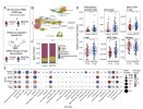
We implemented differential abundance analysis (DESeq2, Materials and Methods) and identified 743 unique features that differed between cases and controls [Benjamini–Hochberg (BH) adjusted P-values (P- adj) threshold of < 0.05 and a log2 fold change cutoff of ±0.5]. Of these, 608 features were elevated in cases, and 135 were elevated in controls. Genes such as IL18R1, CCR7, FCRL5, PRF1, and IFNLR1 were more abundant in cases, whereas genes such as CCL3, TGFB2, CD109, and COL1A1 were more abundant in controls (Fig. 2A).
In addition, we observed changes in abundance of several mtRNA transcripts between cases and controls. Notably, levels of a mitochondrial ribosome assembly factor (MTG1) were significantly increased, while several transcripts (MTND1P23, MT-TL1, MT-TV, MT-TH, MT-TS2, MT-TT, MTATP8P1, MTND6P3, and MTRNR2L3) were significantly decreased. Many of these mtRNA transcripts are inversely correlated with MTG1 in cases (SI Appendix, Fig. S2).
It says the data is available on GEO but the accession (link) is currently marked as private. The analysis code is on GitHub.
 
Last edited:
INFLR1 is a surprising hit—as far as I know, interferon lambda signaling is almost exclusively in epithelial cells, particularly respiratory tract.

[Edit: though I suppose something other than interferon lambda might upregulate expression of that receptor]
 
I don't really understand what is being done here, from the abstract and the figure shown. They seem to be trying to guess which cells are spilling their RNA most in patients and controls? I wonder if it hoild all be explained by activity levels, via circulation times, cell senescence, lack of normal diapedesis into moved tissues etc.

What we need to understand the disease is the specific nature of the abnormal pathways used for something that could cause symptoms. This seems to prejudge what is going to be interesting and end with no specificity?
 
The discussion focuses on developing a diagnostic test for what is assumed to be a valid pathological category. The reality is that nobody should think clinical cohorts as currently defined will be homogeneous in that way. The interest is in why 10% of cases fall outside the normal domain, not that that skew the mean I suspect.

The irony is that clinicians are probably much more skeptical of their diagnostic groupings than the lab workers.
 
The paper mentions a particularly promising model chosen by highest-test AUC across 100 random splits and applies a Youden threshold of 0.65 - is there a potential for selection bias here?
I suppose they did all that selecting in the 70% training set and then tested it in the 30% data that the model hadn't seen yet.
 
They tested RNA outside of the cell which represents a mixture of RNA fragments released from many different cell types all over the body.

Perhaps one of the more interesting findings is that they found no differences in Viral RNA Signatures between ME/CFS patients and controls. In the theory that there is a virus hiding somewhere in the body where it is difficult to measure, these free RNA's might have given a clue about their presence but none was found. The authors caution however, that few of their reads were from solid tissue.
Our cfRNA metagenomic analysis did not detect significant differences in viral RNA burden between phenotypes. Although viral reads mapping to Retroviridae , Herpesviridae , Flaviviridae , and others were detected, their levels were not significantly altered between phenotypes. Nevertheless, given that few of our reads were from solid tissue, we cannot rule out the presence of viral reservoirs whose cfRNA was not captured from the circulation. Most reports of persistent virus in ME/CFS involve tissues and
 
They also found that in their machine learning models, the accuracy was highly variable depending on the predictors that were included.

The main reason for this was thee fraction of platelet-derived cfRNA, which was apparently lower in ME/CFS patients compared to controls. Not sure what this would mean. The authors argue that it cannot be explained by sample collection or test sites differences.
Certain samples, when placed into the test set, were classified correctly in more than 90% of the models, while others were classified correctly in as few as 11%. We found this distribution to be dependent not on a clinical metric, such as phenotype, nor test site, but rather on the fraction of platelet-derived cfRNA (SI Appendix, Fig. S5 ). The relative platelet fraction of each sample was determined by cell type of origin deconvolution. We further showed that this correlation was not due to a difference in sample collection protocols across set sites, but a true biological difference in platelets between cases and controls. This finding supports the notion that ME/CFS patients experience platelet dysfunction at baseline (
 
The model that reached the best fit (in the 30% test samples) was GLMNET Lasso. It reached a maximum accuracy of 77% (84% sensitivity, and
68% specificity, testing AUC-ROC: 0.81 (95% CI: [0.694 to 0.926]).

This is similar to BiomapAI which reported an accuracy of 72.5% and AUC of 0.82 (but which was given a lot more data).

There is some concern that the machine-learning models are picking up on biological patterns that are not very relevant to ME/CFS because there was no correlation between classification scores and symptoms questionnaires (GH stands for SF-36 general health, MFI-20 is a fatigue questionnaire).
1755005295384.png
 
I don't really understand what is being done here, from the abstract and the figure shown. They seem to be trying to guess which cells are spilling their RNA most in patients and controls? I wonder if it hoild all be explained by activity levels, via circulation times, cell senescence, lack of normal diapedesis into moved tissues etc.
I agree it was a confusing step of the analysis. I always thoughts that the interesting part of cfRNA was to be able to easily capture transcripts from non-PBMCs (potentially even from tissues) without having to go into the tissues. If you’re deconvolving transcripts to a PBMC data set, this is basically giving you no new information than you could’ve gotten from a basic PBMC RNA-seq, which they already did several years ago. And you’re just disregarding what transcripts don’t originate from PBMCs.

It’s not like sequencing cfRNA is any more “diagnostically accessible” than sequencing PBMCs, even if there was a strong signature in their findings.

Is there a reason why a higher platelet-poor spin was not used? Notable that the fraction itself strongly influenced classification success - and, also, HBB (haemoglobin beta) was amongst the top LASSO coefficients.
Since the cfRNA sequencing is done on plasma, platelets would already have been filtered out. But the most likely explanation for the findings is platelet rupture during sample processing, which would have caused intracellular mRNA from platelets to enter the plasma fraction at much higher abundance. They said that there was no difference by sample site, though it could have been a difference in how many samples from each group were handled by someone who didn’t notice signs of rupturing.

The other option is that there is some biological difference that makes platelets more likely to rupture in ME/CFS, which would have been interesting to know but it seems they didn't consider. I’m surprised they didn’t mention this in the text—it’s the first question I or any of my colleagues would jump to.
 
Back
Top Bottom