Genetics: FBXL4

Hutan

Moderator
Staff member
DecodeME candidate ME/CFS gene

In DecodeME, we attempted to link GWAS variants to target genes. Here we discuss the top two tiers of predicted linked genes that we are most confident about –‘Tier 1’ and ’Tier 2’.

We defined genes as Tier 1 genes if: (i) they are protein-coding genes, (ii) they have GTEx-v10 expression quantitative trait loci (eQTLs) lying within one of the FUMA-defined ME/CFS-associated intervals, and (iii) their expression and ME/CFS risk are predicted to share a single causal variant with a posterior probability for colocalisation (H4) of at least 75%. For this definition, we disregarded the histone genes in the chr6p22.2 HIST1 cluster, as their sequences and functions are highly redundant (1). This prioritisation step yielded 29 Tier 1 genes.

For the intervals without Tier 1 genes, three Tier 2 genes were defined as the closest protein-coding genes without eQTL association: FBXL4 (chr6q16.1), OLFM4 (chr13q14.3), and CCPG1 (chr15q21.3).

CHROMOSOME 6q

Chr6q contained no Tier 1 genes, but one Tier 2 gene.

The interval also contains a non-protein long noncoding RNA locus (RP11-436D23.1) (25) which contains a miRNA locus (miR-2113) of unknown function. The allele that increases the risk of ME/CFS is associated with increasing RP11-436D23.1 expression in four tissues: amygdala, anterior cingulate cortex, cortex and hippocampus.


********************
FBXL4 (Tier 2)

• Protein: F-box/LRR-repeat protein 4. UniProt. GeneCards.

• Molecular function: Component of the mitochondria-localised SCF-FBXL4 ubiquitin E3 ligase complex. This complex restricts mitophagy by controlling the degradation of BNIP3 and NIX mitophagy receptors (26,27).

• Cellular function: Regulator of mitophagy.

• Link to disease: Mutations in FBXL4 can cause mitochondrial DNA depletion syndrome caused by elevated mitophagy (28).

• Potential relevance to ME/CFS: Reduction in FBXL4 function is associated with impaired mitochondrial respiratory chain deficiency, which has been reported in a sample of people with ME/CFS (29). Lymphoblasts from people with ME/CFS in one study, however, have not been observed to be depleted in mitochondrial DNA (30).

********************

Reference 26
Nguyen-Dien GT, Townsend B, Kulkarni PG, Kozul KL, Ooi SS, Eldershaw DN, et al. PPTC7 antagonizes mitophagy by promoting BNIP3 and NIX degradation via SCFFBXL4. EMBO Rep. 2024 Aug;25(8):3324–47.

Reference 27
Cao Y, Zheng J, Wan H, Sun Y, Fu S, Liu S, et al. A mitochondrial SCF-FBXL4 ubiquitin E3 ligase complex degrades BNIP3 and NIX to restrain mitophagy and prevent mitochondrial disease. EMBO J. 2023 Jul 3;42(13):e113033.

Reference 28
Bonnen PE, Yarham JW, Besse A, Wu P, Faqeih EA, Al-Asmari AM, et al. Mutations in FBXL4 cause mitochondrial encephalopathy and a disorder of mitochondrial DNA maintenance. Am J Hum Genet. 2013 Sep 5;93(3):471–81.

Reference 29
Tomas C, Brown A, Strassheim V, Elson JL, Newton J, Manning P. Cellular bioenergetics is impaired in patients with chronic fatigue syndrome. PLoS One. 2017;12(10):e0186802.

Reference 30
Missailidis D, Annesley SJ, Allan CY, Sanislav O, Lidbury BA, Lewis DP, et al. An Isolated Complex V Inefficiency and Dysregulated Mitochondrial Function in Immortalized Lymphocytes from ME/CFS Patients. Int J Mol Sci. 2020 Feb 6;21(3).
 
the DecodeME study said:
Four of the eight loci (RABGAP1L, FBXL4, OLFM4,CA10) were associated at p < 0.05 with cases ascertained using post-exertional malaise and fatigue in the UK Biobank and the Netherlands biobank Lifelines.
So this was one locus that was replicated.

"OR is the Odds-Ratio of ME/CFS risk in cases versus controls." The OR for FBXL4 is less than 1, so I think that means that there is less chance of ME/CFS in people who have the variant i.e. the variant is protective.

Screenshot 2025-08-07 at 9.14.54 pm.png

I think this being a Tier 2 variant, it means that we don't know what the effect of the variant is? Does it up or down regulate FBXL4 and the mitophagy restriction?

FBXL4 regulates mitophagy, which is a process where surplus, aged or damaged mitochondria are degraded.

When FBXL4 is absent, the levels of BNIP3 and NIX raise and hyperactive mitophagy results. Loss-of-function mutations in FBXL4 are known to cause mitochondrial DNA depletion syndrome 13.

In DecodeME, the FBXL4 variant that was associated with ME/CFS was rs97984426. I couldn't find information on whether it would lead to icreased or decreased mitophagy.

In this study of mitochondrial DNA it was reported that "ME/CFS patients had an excess of individuals without a mildly deleterious population variant".
https://www.nature.com/articles/s41598-019-39060-1

Fewer deleterious mtDNA variants would suggest higher mitophagy is occurring. The question is how could this contribute to ME/CFS?
 
I wonder what this protein is regulated by - what changes it responds to. That might provide a clue.

In the same section of CP's "Candidate" document, I am puzzling over this nearby miR-2113 locus:

"The interval also contains a non-protein long noncoding RNA locus (RP11-436D23.1) (25) which contains a miRNA locus (miR-2113) of unknown function. The allele that increases the risk of ME/CFS is associated with increasing RP11-436D23.1 expression in four tissues: amygdala, anterior cingulate cortex, cortex and hippocampus."
 
Have we seen this gene before in ME/CFS research?

Or am I just thinking of the Crawley pediatric LC study?


Here’s something:

That's a really cool resource, thanks. For reference here is the paper the genes are from: Genetic Risk Factors for ME/CFS Identified using Combinatorial Analysis (Das et al, 2022, J Transl Med)

And here are the 14 genes they found, linked to their Protein Atlas cell type page (GC links to GeneCards page). I added where these proteins seem to be concentrated, just from a visual impression.
S100PBP - GC (glial, spermatocytes/spermatogonia)
ATP9A - GC (neuronal, glial)
KCNB1 - GC (neuronal)
CLOCK - GC (generally equal among cell types)
SLC15A4 - GC (dendritic)
TMEM232 - GC (excitatory/inhibitory neurons, glial, germ, ciliated)
GPC5 - GC (astrocytes)
PHACTR2 - GC (generally equal among cell types)
AKAP1 - GC (late spermatids)
USP6NL - GC (glial - mostly microglia)
CDON - GC (muller glia, excitatory/inhibitory neurons, mesothelial)
INSR - GC (generally equal among cell types)
SLC6A11 - GC (mainly astrocytes, but also other glial and neuronal cells)
SULF2 - GC (oligodendrocyte precursor, granulosa, endometrial stromal, maybe dendritic)

Edit: Also, just checked and there is no overlap between these genes and the 115 Zhang genes. The highest ranked of these is AKAP1 at position 1159 in the full Zhang list of 17759 genes.
 
Molecular function: Component of the mitochondria-localised SCF-FBXL4 ubiquitin E3 ligase complex. This complex restricts mitophagy by controlling the degradation of BNIP3 and NIX mitophagy receptors (26,27).

Reference 27
Cao Y, Zheng J, Wan H, Sun Y, Fu S, Liu S, et al. A mitochondrial SCF-FBXL4 ubiquitin E3 ligase complex degrades BNIP3 and NIX to restrain mitophagy and prevent mitochondrial disease. EMBO J. 2023 Jul 3;42(13):e113033.

[27] posted on thread A mitochondrial SCF‐FBXL4 ubiquitin E3 ligase complex degrades BNIP3 and NIX to restrain mitophagy and prevent mitochondrial disease (2023)
 
Commenting in the main DecodeME thread:
I know some of our members have diagnosed issues with iron, and infections can change iron homeostasis.

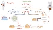
BNIP3 has links to HIF-1⍺, iron metabolism and the proteasome. This paper suggests a bi-directional upregulation between BNIP3 and HIF-1⍺ (in a melanoma model).

Screenshot 2025-08-09 at 3.33.23 PM copy.jpg

BNIP3 promotes HIF‐1α‐driven melanoma growth by curbing intracellular iron homeostasis (2021, The EMBO Journal)

BNIP3 is a mitophagy receptor with context‐dependent roles in cancer, but whether and how it modulates melanoma growth in vivo remains unknown. Here, we found that elevated BNIP3 levels correlated with poorer melanoma patient’s survival and depletion of BNIP3 in B16‐F10 melanoma cells compromised tumor growth in vivo. BNIP3 depletion halted mitophagy and enforced a PHD2‐mediated downregulation of HIF‐1α and its glycolytic program both in vitro and in vivo.

Mechanistically, we found that BNIP3‐deprived melanoma cells displayed increased intracellular iron levels caused by heightened NCOA4‐mediated ferritinophagy, which fostered PHD2‐mediated HIF‐1α destabilization. These effects were not phenocopied by ATG5 or NIX silencing. Restoring HIF‐1α levels in BNIP3‐depleted melanoma cells rescued their metabolic phenotype and tumor growth in vivo, but did not affect NCOA4 turnover, underscoring that these BNIP3 effects are not secondary to HIF‐1α.

These results unravel an unexpected role of BNIP3 as upstream regulator of the pro‐tumorigenic HIF‐1α glycolytic program in melanoma cells.

• High BNIP3 expression in melanoma patients correlates with poor survival.

• BNIP3 deficiency in mice reduces melanoma growth.

• BNIP3‐depleted melanoma cells show reduced HIF‐1α protein levels, glycolysis defects and exacerbated lysosomal turnover of the ferritinophagy receptor NCOA4.

• Elevated intracellular iron in BNIP3‐deprived cells promotes PHD2‐mediated degradation of HIF‐1α.

• A HIF‐1α hydroxylation‐mutant rescues tumor growth potential of BNIP3‐silenced cells.

Web | PDF | The EMBO Journal | Open Access

We propose a model in which BNIP3, due to its ability to regulate the intracellular availability of iron by directly controlling NCOA4-mediated ferritinophagy, maintains HIF-1α-driven glycolytic program and establishes a feedforward BNIP3-HIF-1α axis that fosters melanoma growth. This bidirectional loop between BNIP3 and HIF-1α is an unexpected finding of this study, given that BNIP3 is a hypoxia-responsive gene thought to operate as a downstream target of HIF-1α rather than being a positive regulator of this transcription factor.

 
Copy of @chillier post to add info to this thread from main thread of the paper for this gene:

FBXL4 can cause mitochondrial DNA depletion syndrome caused by elevated mitophagy.

Loss of function causes excess culling of mitochondria leading to mitochondrial DNA depletion syndrome or mitochondrial encephalopathy. This reminded me of something from Josh Dibble's 2020 review about mitochondrial genetics:

Independent studies confirm that clinically proven mitochondrial DNA (mtDNA) variants do not commonly explain ME/CFS (26–28). People with ME/CFS, however, appear more likely to carry mtDNA that lacks even mildly deleterious variants (28), a finding whose implications require further investigation.

I wonder if an FBXL4 mediated oversensitivity to removing slightly mutated mitochondria could explain that. It appears that FBXL4 loss is deleterious to mitochondrial function - from FBXL4's OMIM page:

Patient muscle homogenates or isolated mitochondria showed variably decreased activities of the mitochondrial respiratory chain complexes as well as decreased mtDNA content. Cultured skin fibroblasts had reduced maximal oxygen consumption rate and increased fragmentation of the mitochondrial network. At least 1 patient cell line studied showed a significant reduction of the mitochondrial membrane potential. These defects could be rescued by expression of wildtype FBXL4 in patient cells. The findings indicated that FBXL4 is necessary for the homeostasis of mitochondrial bioenergetics.
 
Copy of post from @DMissa of main thread of the paper for this gene:

Patient muscle homogenates or isolated mitochondria showed variably decreased activities of the mitochondrial respiratory chain complexes as well as decreased mtDNA content. Cultured skin fibroblasts had reduced maximal oxygen consumption rate and increased fragmentation of the mitochondrial network. At least 1 patient cell line studied showed a significant reduction of the mitochondrial membrane potential. These defects could be rescued by expression of wildtype FBXL4 in patient cells. The findings indicated that FBXL4 is necessary for the homeostasis of mitochondrial bioenergetics.

We are already looking at this stuff so whether this effect holds up or not (presumably more subtly than this, if so) it will actually come to light as a matter of time.

I was intending to vomit out my encyclopaedic knowledge of the hodgepodge of mitochondrial findings available to provide context to how we interpret this gene coming up (and expected effects of issues with it) but, really, since we have little that is consistent or replicated I actually think it wouldn't really be good to make any major predictions. tldr is some parts of the above cited phenotype have manifested in different studies but variably between sample types and usually with small effect sizes, hard to draw anything definitive. eg: membrane potential reduction.

What's probably more interesting than going back to what we "know" about mitochondrial studies in ME/CFS is the sort of orthogonal line of enquiry this invites. As far as I am aware mitophagy hasn't really been looked at in published work on ME/CFS. Instead much of the research involving mitochondria has focused on respiratory activity, sometimes on morphology. While morphology is tricky to interpret from particularly older literature where methods were more subjective, there have actually been generally striking differences reported, and in several tissues. It doesn't come up in the discussion often because of the difficulty of interpretation and age of some of the work. But changes in morphology are very clear, direct consequences of changes in how mitophagy is working so if there's anything to that, it's a point in favour.

Anyway. Looking forwards:

I am actually quite surprised that something like this came up (and pleasantly, because it's plausible and interesting). Mitophagy is an interesting angle and issues with it might make more sense in terms of how I imagine things actually happening. Issues with mitochondrial biogenesis, quality control, dynamics and whatever else can very easily elicit stress signalling responses that could plausibly drive many of the issues we have been recently suspecting here on s4me. A mitophagy issue would match the clinical picture much more than the commonly regurgitated blanket "energy production issue". You can have consequences for sensitivity and activation of immune cells, entanglement with ER issues more generally and the list goes on. It's very fertile ground. And to somebody else who I know will read this, wink wink nudge nudge, I won't steal your thunder.

Not as a plug but as a point of optimism and excitement, our lab is particularly well placed to look at many components of this. We are already doing complementary things and we have some unique means to study these processes that aren't commercially available. Hopefully we can add some clarity to whether there are functional consequences here that align with clinical presentation.

I'm starting to design some projects, not just specifically for this gene and mitochondria, but there's my 2c for anyone that wants the quick summary of where we can go with this particular gene.

Apologies if this isn't clear or organised, I'm unwell today but I couldn't just sleep on today's news.

The point is: mitophagy, and more generally the consequences for a cell should mitochondria be forming or breaking up, being cleaned up or talking to other organelles in a crappy way, is a plausible and compelling line of investigation (in my view moreso than a lot of the mitochondrial ideas that are typically memed to death, usually accompanying vague evocations of "energy" and/or "fatigue" - yes I have also been guilty of this).
 
Back
Top Bottom