Loss of CRH neurons and other neural changes in ME/CFS autopsy study - University of Amsterdam

These results are not definite and sometimes contested but it seems longer illness duration has a trend toward more severity. Which I would in turn expect for people at the end of their lives along with comorbidity.
For a start, the unremarkable fact of the additional burdens of normal ageing on top of serious chronic and very badly managed disease need to be factored in. If you are already barely functional it does not take much extra burden to crash the whole show.
I think those studies have all kinds of potential issues with sampling bias. It’s a sign of how badly we’ve been treated that we don’t have good data on this yet.
Indeed. The lack of decent epidemiological data after decades of 'study' tells you everything you need to know about how seriously this has not been taken (at least until recently). Should have been among the first major studies to be done.
 
There is an official government funded program for ME CFS research with this brain bank - under the supervision of the NMCB-consortium.
It's a terrific achievement to get that program in place.


I think the relevance is mainly in the fact that CSF cortisol was measured in the NIH study and not found to be abnormal. That makes the result of abnormal CSF cortisol here less likely to be true, and contributes to the likelihood that something has gone wrong with the sampling that affected the integrity of the CRH cells.
Not sure. I think the possibility that the CRH cell findings are artifactual is probably an independent issue relating to histological methodology (not particularly sampling).
A problem with the cortisol measurement and a problem with the CRH cell findings could be separate issues. But finding one problem does tend to make it more likely that there is another, regardless of whether the problems are directly linked.

But, I hear @Arfmeister and others suggesting this team are good at what they do. I guess the published paper will go some way to answering our questions.
 
Not that you necessarily need this clarified, but just to make this thread as clear as possible: the people in this study were autopsied because they signed up to donate to the brain bank's ME/CFS program before they passed. (At least that is my understanding.)

I don't think the researchers have released any info yet about the patients, such as if they passed due to euthanasia, or how severe their ME/CFS was. (But please let me know if I just missed it.) It's true more severe patients might be more likely to sign up for a study like this, and also true that there are far more mild patients, so hard to know how the math works out there.

The brain bank says that their patient samples are "anonymized". I don't know if that just means that their names are not included, or if things like cause of death are going to be impossible to find out.
Yes, it's probably also worth mentioning that a standard post mortem is very different from what a brain bank does/can do. There seems to be some confusion about this - the idea that ME/CFS is not 'a classic neurological disease' because post mortems have been largely unremarkable has always been bogus and extremely superficial/premature. In fact, it's such a ridiculous idea that it's difficult to not see it as a political ploy.

Some of the more standard post mortems in the past have actually found stuff that 'made sense' like the DRG findings, but what little has been published or is known about it has never been systematized and/or is of low quality.
 
Last edited:
We might have a problem that a brain bank may not look at spinal cord. There was no mention of dorsal root ganglia in the CRH neuron study as far as I know and maybe they didn't look.
Absolutely, it would be a huge mistake to not include the spinal cord - I am not sure whether that's feasible within the brain bank framework. @Grigor do you have any knowledge of this and/or had prior contact with them and uncomplicated ways to ask them about it?
 
I no longer have access to mapMECFS, the platform for sharing raw data for the deep phenotyping study. But I previously posted about doing statistical tests on all 435 CSF metabolites, and cortisol was in fact one of the chemicals that was in the data.

And in another post where I posted the results for the top 40 most significant metabolites out of these 435 based on Mann-Whitney testing, cortisol was not one of those with high significance.
I was waiting to get a reply from someone at mapMECFS to see if it would be okay for me to still post the data I previously analyzed, and I was told I could.

I see it's already been mostly discussed at length, but they also suggested I note that there may be reasons for the differences, such as duration of illness, and the fact that cortisol fluctuates throughout the day, and possibly even changes significantly after death.

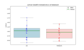
But here is the cortisol CSF data from the deep phenotyping study. There appears to be virtually no difference between the groups.

One note is that I have some concerns that there might have been some sort of issue with the CSF metabolomics part of the deep phenotyping study, as nearly all of the 435 metabolites (88%) were lower in ME/CFS. I posted about it on PubPeer. Although if metabolites were biased lower, presumably that would make a true low cortisol finding look even more significant, not less.

Edit: When comparing these groups with a Mann-Whitney test, p=0.467.
 
Last edited:
Bumped into this old study that might be relevant to the CRH-autopsy findings.

Abnormalities in response to vasopressin infusion in chronic fatigue syndrome​

M Altemus 1, J K Dale, D Michelson, M A Demitrack, P W Gold, S E Straus

Abstract​

Several neuroendocrine studies have suggested hypoactivation of the hypothalamic-pituitary-adrenal axis in chronic fatigue syndrome. One possible determinant of this neuroendocrine abnormality, as well as the primary symptom of fatigue, is reduced hypothalamic secretion of corticotropin-releasing hormone (CRH). Because CRH and vasopressin secreted from the hypothalamus act synergistically at the pituitary to activate ACTH secretion, the ACTH response to peripheral infusion of vasopressin can provide an indirect measure of hypothalamic CRH secretion. We measured the ACTH and cortisol response to a one hour infusion of arginine vasopressin in 19 patients with chronic fatigue syndrome and 19 age and sex matched healthy volunteers. Patients with chronic fatigue syndrome had a reduced ACTH response to the vasopressin infusion and a more rapid cortisol response to the infusion. These results provide further evidence of reduced hypothalamic CRH secretion in patients with chronic fatigue syndrome.
Abnormalities in response to vasopressin infusion in chronic fatigue syndrome - PubMed
 
Bumped into this old study that might be relevant to the CRH-autopsy findings.
A relatively small study, with great overlap between the CFS and control groups. I don't think the study provides convincing evidence that there is anything amiss with the response to the infusion of vasopressin.

Of course, the small size, dropouts and possible problems with selection in that study means that we can't completely rule out the possibility of reduced CRH secretion in a subset of people with ME/CFS.
 
Cross-posting my question on the Prusty et al. brain autopsy study thread here for visibility, since this thread is newer and more active.
...
Does the success of the Netherlands Brain Bank (NBB) in establishing a source for brain tissue (used in the "Loss of CRH Neurons" study, with apparently more to come) create an opportunity for replicating this HHV6/EBV study? Does anyone have the connections within the research community to facilitate a conversation about that possibility?
...
(If I put in in the wrong place to begin with, moderators feel free to move the original - still learning forum norms).
 
I was waiting to get a reply from someone at mapMECFS to see if it would be okay for me to still post the data I previously analyzed, and I was told I could.

I see it's already been mostly discussed at length, but they also suggested I note that there may be reasons for the differences, such as duration of illness, and the fact that cortisol fluctuates throughout the day, and possibly even changes significantly after death.

But here is the cortisol CSF data from the deep phenotyping study. There appears to be virtually no difference between the groups.


One note is that I have some concerns that there might have been some sort of issue with the CSF metabolomics part of the deep phenotyping study, as nearly all of the 435 metabolites (88%) were lower in ME/CFS. I posted about it on PubPeer. Although if metabolites were biased lower, presumably that would make a true low cortisol finding look even more significant, not less.

Edit: When comparing these groups with a Mann-Whitney test, p=0.467.
I often wonder how much these CSF measurements are influenced by the fact that a doctor presents patients with a long, scary needle that he plans to stick into their spine. Probably even worse for adrenaline.

That might explain the wide ranges that Hutan found.

Nevertheless, I suppose it shows that pwME can still release the same amount of cortisol as healthy controls under these conditions.
 
I often wonder how much these CSF measurements are influenced by the fact that a doctor presents patients with a long, scary needle that he plans to stick into their spine. Probably even worse for adrenaline.

That might explain the wide ranges that Hutan found.

Nevertheless, I suppose it shows that pwME can still release the same amount of cortisol as healthy controls under these conditions.
I was in an ME/CFS study several years back where a lumbar puncture was required. I'd had a couple for other studies, so I wasn't overly concerned. But it took them well over an hour with that long needle embedded in my spine to withdraw enough fluid. They even manipulated the gurney they had me on to see if gravity could help the process. I guarantee that my adrenaline was flowing.

If I recall correctly, most of my values, including cortisol, were unremarkable.
 
I was in an ME/CFS study several years back where a lumbar puncture was required. I'd had a couple for other studies, so I wasn't overly concerned. But it took them well over an hour with that long needle embedded in my spine to withdraw enough fluid. They even manipulated the gurney they had me on to see if gravity could help the process. I guarantee that my adrenaline was flowing.

If I recall correctly, most of my values, including cortisol, were unremarkable.
Thank you for participating in that study and sharing how your cortisol was (not) affected. I’m grateful to every patient who puts themselves through lumbar punctures for research. I hate needles too much to ever be able to do it myself.
 
This German article discusses the brain autopsy Study Netherlands.
Most interesting part is that apparently in the US 100 brain samples from people with Long COVID are currently being studied.

preliminary findings from Dutch researchers who examined the brains of people who had died with severe ME/CFS. According to the report, these are the first systematic brain autopsy studies specifically focused on ME/CFS and they point toward abnormalities in the hypothalamus, a brain region that regulates stress, energy balance, sleep, body temperature, and hormone production.
Main findings
* Researchers examined the brains of seven people with severe ME/CFS, comparing them with carefully selected controls. The tissue came from the Netherlands Brain Bank’s dedicated ME/CFS brain donation program.
* The most striking observation was a marked reduction in neurons that produce corticotropin-releasing hormone (CRH) in the hypothalamus. Some cases reportedly had very few CRH-producing neurons remaining. Other neuron types, such as those producing vasopressin and oxytocin, appeared normal or even increased.
* The researchers also found abnormalities in the pituitary gland, including reduced expression of hormone receptors and precursor hormones. Together, these findings suggest a severely impaired hypothalamic-pituitary-adrenal (HPA) axis, the body’s central stress-response system.

Possible cause
The researchers do not claim to know what caused these brain changes. However, they discuss the possibility that chronic neuroinflammation may progressively damage stress-regulating brain regions, particularly the CRH neurons of the hypothalamus. This remains a hypothesis rather than a proven mechanism.

Treatment implications
If the brain’s stress-regulation system is structurally damaged, simply replacing cortisol with low-dose steroids may not fully correct the underlying problem. Instead, future therapies might need to target:
* Neuroinflammation
* Immune dysregulation
* Central nervous system dysfunction
These approaches are still experimental and are not established treatments.

Relevance to Long COVID
*The article notes that several 100 brain samples from people with Long COVID are currently being studied through the U.S. RECOVER program. If similar abnormalities are found, it could indicate shared mechanisms between ME/CFS and Long COVID.*

 
Last edited:
Back
Top Bottom