[Preprint] Post-COVID syndrome associated with capillary alterations, macrophage infiltration & distinct transcriptomic signatures in skeletal muscles

SNT Gatchaman

Senior Member (Voting Rights)
Staff member
Post-COVID syndrome is associated with capillary alterations, macrophage infiltration and distinct transcriptomic signatures in skeletal muscles
Tom Aschman, Emanuel Wyler, Oliver Baum, Andreas Hentschel, Franziska Legler, Corinna Preusse, Lil Meyer-Arndt, Ivana Buettnerova, Alexandra Foerster, Derya Cengiz, Luiz Gustavo Teixeira Alves, Julia Schneider, Claudia Kedor, Rebekka Rust, Judith Bellmann-Strobl, Aminaa Sanchin, Peter Vajkoczy, Hans-Hilmar Goebel, Markus Landthaler, Victor Corman, Andreas Roos, Frank L. Heppner, Helena Radbruch, Friedemann Paul, Carmen Scheibenbogen, Werner Stenzel, Nora F. Dengler

The SARS-CoV-2 pandemic not only resulted in millions of acute infections worldwide, but also caused innumerable cases of post-infectious syndromes, colloquially referred to as long COVID. Due to the heterogeneous nature of symptoms and scarcity of available tissue samples, little is known about the underlying mechanisms.

We present an in-depth analysis of skeletal muscle biopsies obtained from eleven patients suffering from enduring fatigue and post-exertional malaise after an infection with SARS-CoV-2. Compared to two independent historical control cohorts, patients with post-COVID exertion intolerance had fewer capillaries, thicker capillary basement membranes and increased numbers of CD169+ macrophages. SARS-CoV-2 RNA could not be detected in the muscle tissues, but transcriptomic analysis revealed distinct gene signatures compared to the two control cohorts, indicating immune dysregulations and altered metabolic pathways.

We hypothesize that the initial viral infection may have caused immune-mediated structural changes of the microvasculature, potentially explaining the exercise-dependent fatigue and muscle pain.

Link
 
Biopsies were taken in average one year after initial infection and were compared to historical control samples, consisting of histologically normal samples as well as samples with a selective atrophy of type-2b muscle fibers. The latter is a non-specific finding associated with muscle disuse and immobility.

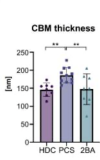
While patients with PCS did not show overt signs of myositis, increased numbers of CD169 + macrophages were evident. The capillary-to-fiber ratio was decreased and ultrastructural analysis revealed a capillary basement membrane enlargement. SARS-CoV-2-specific RNA was not detected [...] transcriptomic analysis showed distinct transcriptomic profiles in most of the PCS samples compared to the controls, with increased expressions of genes related to immune system regulation, angiogenesis and extracellular matrix remodeling, and decreased expressions of genes related to metabolic processes and mitochondrial activity.

Numbers of capillaries per muscle fiber (C/F) were quantified by assessing at least 10 images of semithin cross-sections of all included muscle specimens at 200x magnification. This revealed a significant decrease in the PCS cohort when compared to the two historical control cohorts (PCS vs. HDC: mean difference 0.32, p=0.007; PCS vs. 2BA: mean difference 0.30, p=0.013).

Comparison of mean cross-sectional fiber area (MCSFA) revealed a significant decrease in the PCS cohort compared to the 2BA cohort (mean difference 1508 µm2 , p=0.009) whereas no significant difference was present when compared to the 2BA cohort. The reduced MCSFA explains why there was no significant difference of the capillary density between the groups. In fact, capillary density represents the absolute number of capillaries per mm 2 and due to a decreased MCSFA, more fibers and more capillaries will be observed in one field of vision. Therefore, interpretation of the C/F is more conclusive.

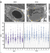
In one patient (PCS-4), massive structural damage was observed in all 23 photographed capillary profiles. Endothelial cells were almost completely degenerated, resulting in debris-containing empty capillary tubes also called string vessels (=acellular capillary remnants). Such severe morphological alterations were not found in any other patient. Immunohistochemistry revealed the presence of many large CD169 + macrophages in close vicinity to the capillaries.

Of note, the patient in which we identified massive structural capillary damage (PCS-4) constituted a clear outlier with much higher expression levels of mostly immune system related genes.
 
Looks like a carefully done study, with the authors having a decent understanding of ME/CFS.

Quantification of immune cells revealed a significant increase of CD68+ (HDC vs. PCS: mean of 32 [SD 12] versus 58 [SD 16] cells/10HPF, p=0.002; 2BA vs. PCS: mean of 34 [SD 15] versus 58 [SD 16] cells/10HPF, p=0.005) and CD169+ (HDC vs. PCS: mean of 17 [SD 10] versus 41 [SD 13] cells/10HPF, p= 0.0004; 2BA vs. PCS: mean of 22 [SD 11] versus 41 [SD 13] cells/10HPF, p= 0.004) macrophages. Numbers of CD45+ cells, CD8+ T-cells and CD206+ macrophages were not significantly different between the three groups (Fig. 2B-C).

The following is some googling, to understand what CD68+ and CD169+ macrophages do, and to see if they have been mentioned in connection with ME/CFS before:

About CD68+
"CD68 is a heavily glycosylated glycoprotein that is highly expressed in macrophages and other mononuclear phagocytes. Traditionally, CD68 is exploited as a valuable cytochemical marker to immunostain monocyte/macrophages in the histochemical analysis of inflamed tissues, tumor tissues, and other immunohistopathological applications."

There's a paper that notes that the presence of antibodies to Covid spike protein results in CD68+ cells internalising the SARS (1) virus and virus particles.

There's a French researcher, Authier, who looks to have been finding and reporting (for years) high levels of macrophages in muscle tissue and noting that most of the patients with this had chronic fatigue. He was calling the problem macrophagic myofasciitis, and suggesting that it was either a cause of CFS or should be an exclusion criterion. I haven't looked at that hard, there's mention of aluminium adjuvants from vaccines, perhaps he wasn't getting everything right. Nevertheless, it is interesting that someone was finding that an increase of macrophages in muscle tissue was associated with CFS-like symptoms.

Ultraendurance athletes were found to have an increase of CD68+ in muscle tissue of a similar magnitude to that found here (i.e a doubling) a day after 24 hours of ongoing exercise.


About CD169+
The authors of this 2023 Aschmann et al paper seem quite impressed by the increase in CD169+ levels. They said:
The mere fact that the number of CD169+ macrophages is increased in mildly altered skeletal muscle tissue is remarkable. CD169+ macrophages are increased in idiopathic inflammatory myopathies (54), indicating a prominent role in type I Interferon-related immune processes (55), and recent studies emphasized their highly specific functional programmes and important roles as border-associated cells at blood vessel/parenchymal interfaces (56, 57). CD169+ macrophages have further been implicated in antiviral defense, being the primary cell infected and able to capture viral particles in the blood and subsequently presenting them to B cells (58). As numbers of circulating CD169+ monocytes are increased in acute stages of mild COVID-19 (59), we hypothesize that they could play a key role at the muscle/capillary interface in our cohort of patients with PCS.

There's a good recent review of CD169+ here:
Functions of CD169 positive macrophages in human diseases (Review), 2020

CD169 macrophages internalise viruses and even allow them to replicate, in order to be able to present bits of them to immune cells, activating an immune response.

CD169-mediated restrictive SARS-CoV-2 infection of macrophages induces pro-inflammatory responses 2022
CD169-mediated SARS-CoV-2 entry in macrophages resulted in expression of viral genomic and subgenomic RNAs with minimal viral protein expression and no infectious viral particle release, suggesting a post-entry restriction of the SARS-CoV-2 replication cycle. Intriguingly this post-entry replication block was alleviated by exogenous ACE2 expression in macrophages.


It seems that it is possible for bacteria to survive and replicate within a CD169+ expressing cell, potentially creating a reservoir for septicaemia
Intracellular replication of Streptococcus pneumoniae inside splenic macrophages serves as a reservoir for septicaemia.
occasional intracellular replication of bacteria that are taken up by a subset of CD169+ splenic macrophages. In this model, proliferation of these sequestered bacteria provides a reservoir for dissemination of pneumococci into the bloodstream, as demonstrated by its prevention using an anti-CD169 monoclonal antibody treatment. Intracellular replication of pneumococci within CD169+ splenic macrophages was also observed in an ex vivo porcine spleen, where the microanatomy is comparable with humans.


There's this 2011 study by Andrew Lloyd et al where they looked at gene expression in peripheral blood. One of the genes identified was associated with CD169
Peripheral Blood Gene Expression in Postinfective Fatigue Syndrome Following From Three Different Triggering Infections
Three of the genes associated with neurocognitive disturbance in the EBV group had an adjusted significance level of <.05, including glutamate decarboxylase-2 (GAD2), sialoadhesion-1 (CD169; SIGLEC-1), and serine/threonine/tyrosine interacting protein (STYX).
I found it interesting to skim read that one. It comes from a time when perhaps Lloyd had been a bit more open-minded about there being a biological cause. The study, which was hardly extensive, with a total of 18 samples from 3 different triggering diseases and which demanded that genes be found that were expressed in all three of the groups with different disease triggers, led Lloyd and his fellow authors to conclude
The major finding reported here with the use of this comprehensive approach was that no genes were consistently associated with the illness....
A growing body of evidence points to the central nervous system as the likely site of the pathophysiological disturbance underpinning CFS [5, 6, 46]. ... The data presented here suggest that further investigation of the peripheral blood transcriptome is not warranted.
It would be bleakly funny if the expression of CD169/SIGLEC-1 turned out to be relevant to ME/CFS.
 
Last edited:
We hypothesize that the initial viral infection may have caused immune-mediated structural changes of the microvasculature, potentially explaining the exercise-dependent fatigue and muscle pain.

Although an interesting study with relevance to ME, this hypothesis seems to suggest that Long Covid with PEM results from a fixed term/one off event resulting in structural changes during the acute viral infection. Does this then imply after the causal infection the ongoing condition should be stable or gradually remitting as the body attempts to repair itself?

Would ME and I suspect Long Covid meeting the diagnostic criteria for ME be better characterised as an ongoing process?

Certainly a characteristic feature of ME is its variability both within and between individuals. My initial experience of ME could fit in with the idea of a one off onset event triggered by an acute infection, EBV/glandular fever, which though fluctuating displayed overall improvement over a number of years and again my first major relapse was associated with a bout of seasonal flue with subsequent overall improvement albeit slower again over a number of years are not inconsistent with this hypothesis. However in contrast my next rapid onset major relapse began as PEM the day after a twelve mile hike followed by even slower improvement over a number of years, and my current severe impairment involved more gradual deterioration over a number of years with no easily identified trigger events. I don’t think the variation in individuals’ condition is explainable, without out recourse to other factors, as a one off acute event, nor is the different courses of the condition seen in different sufferers, ranging from gradually improving, to stable, to relapsing/remitting to ongoing deterioration.

It would be interesting to understand what changes in the microvasculature occur within individuals over time and if they correlate with the severity of symptoms over time.
 
I read it more that the virus sets up the milieu whereby immune dysregulation has a number of deleterious downstream events, esp on the endothelium. I would expect onset to be slow and inexorable, but at the same time homeostatic mechanisms that can partly compensate could keep symptoms variable in degree and location, and sometimes under control in the initial stages.

Our findings suggest persistent local immune responses in subsets of patients with PCS even one year after initial infection, which in the absence of evidence for an unresolved infection and the presence of autoantibodies in some individuals from our cohort, may point towards immune system dysregulations or an autoreactivity, consistent with multiple observations in patients with acute and post-acute COVID-19.

For me at the start, I would have weeks of postural tachycardia which would then subside, with two to three weeks of normality. This cycled for a couple of months before symptoms bedded in, and then proceeded to worsen a few months later.
 
CD68 is a general macrophage marker. CD169 probably equates to active scavenger macrophages with antigen-presenting capabilities. I am not sure that it tells us more than that macrophages have recently been called in. it may do.

I would be interested to know what these muscles look like stained for CD55 or CD16. As I have mentioned before, different tissues carry different signalling molecules adsorbed on to fibrillin fibres in their elastic networks. These molecules are normally thought of as functioning on cell surfaces but seem to get painted on to the matrix network as well, where they may provide a 'response memory' for the tissue. I can imagine that after an acute infection these signalling proteins may get left on fibrillin for months, possibly acting as a sump for immune complex binding and low grade cytokine activation.

Looking for cells is of interest but always leaves the question as to whether they are causing a problem or responding to one. Finding changes in matrix might help sort that out.
 
I think this is the first time I have seen an academic paper not written by patients that correctly frames it as exertion intolerance, rather than about exercise. My memory being bad, no more than a handful for sure. Kudos for that alone, it's rare to pay attention to the right details, even after 3 years.

Bit funny to speak of localized immune responses talking about one of the two organs that span every single part of the body. Not saying this is the case but it'd be kind of funny if, basically: good news, we found the problem in one organ, bad news, that organ is everywhere, and I do mean literally everywhere. It's localized everywhere, basically. But at least it restricts the search space anyway.

Is there good expertise in vascularity that isn't simply a subset of cardiology and still views the heart as the only interesting part? Seems like an area that isn't especially large or exciting.
 
Am I correct in thinking that this study provides experimental support for Fluge and Mella’s proposed pathological mechanism of endothelial dysfunction? They published a study on it this year.

In any case, this study seems important on its own.
 
Back
Top Bottom