Sex differences and immune correlates of Long COVID development, persistence, and resolution, 2024, Hamlin et al.

Discussion in 'Long Covid research' started by SNT Gatchaman, Jun 20, 2024.

  1. SNT Gatchaman

    SNT Gatchaman Senior Member (Voting Rights)

    Messages:
    5,923
    Location:
    Aotearoa New Zealand
    Sex differences and immune correlates of Long COVID development, persistence, and resolution
    Rebecca E Hamlin; Shaun M Pienkos; Leslie Chan; Mikayla A Stabile; Kassandra Pinedo; Mallika Rao; Philip Grant; Hector Bonilla; Marisa Holubar; Upinder Singh; Karen B Jacobson; Prasanna Jagannathan; Yvonne Maldonado; Susan P Holmes; Aruna Subramanian; Catherine A Blish

    Sex differences have been observed in acute COVID-19 and Long COVID (LC) outcomes, with greater disease severity and mortality during acute infection in males and a greater proportion of females developing LC. We hypothesized that sex-specific immune dysregulation contributes to the pathogenesis of LC.

    To investigate the immunologic underpinnings of LC development and persistence, we used single-cell transcriptomics, single-cell proteomics, and plasma proteomics on blood samples obtained during acute SARS-CoV-2 infection and at 3 and 12 months post-infection in a cohort of 45 patients who either developed LC or recovered.

    Several sex-specific immune pathways were associated with LC. Specifically, males who would develop LC at 3 months had widespread increases in TGF-β signaling during acute infection in proliferating NK cells. Females who would develop LC demonstrated increased expression of XIST, an RNA gene implicated in autoimmunity, and increased IL1 signaling in monocytes at 12 months post infection.

    Several immune features of LC were also conserved across sexes. Both males and females with LC had reduced co-stimulatory signaling from monocytes and broad upregulation of NF-κB transcription factors. In both sexes, those with persistent LC demonstrated increased LAG3, a marker of T cell exhaustion, reduced ETS1 transcription factor expression across lymphocyte subsets, and elevated intracellular IL-4 levels in T cell subsets, suggesting that ETS1 alterations may drive an aberrantly elevated Th2-like response in LC.

    Altogether, this study describes multiple innate and adaptive immune correlates of LC, some of which differ by sex, and offers insights toward the pursuit of tailored therapeutics.


    Link | PDF (Preprint: BioRxiv) [Open Access]
     
  2. SNT Gatchaman

    SNT Gatchaman Senior Member (Voting Rights)

    Messages:
    5,923
    Location:
    Aotearoa New Zealand
    ETS1 (GeneCards)

    Corroborated by Blood transcriptomic analyses reveal persistent SARS-CoV-2 RNA and candidate biomarkers in post-COVID-19 condition (2024, The Lancet Microbe) —

     
  3. Hutan

    Hutan Moderator Staff Member

    Messages:
    29,697
    Location:
    Aotearoa New Zealand
    Genecards mentioned a link with Lupus.
    Googling ETS1 and Lupus:
    The role of the transcription factor Ets1 in lupus and other autoimmune diseases, 2016

     
  4. Hutan

    Hutan Moderator Staff Member

    Messages:
    29,697
    Location:
    Aotearoa New Zealand
    There's a lot in this study - it's a bit overwhelming in terms of sorting out what is going on and what is worth looking at .

    Darker red means a stronger association between higher NFKB1 expression and Long covid at 12 months - for various cell types.
    It's notable that there is no blue, which would indicate lower NFKB1 expression.
    For males and females combined

    Screen Shot 2024-06-20 at 10.25.32 pm.png
     
  5. Hutan

    Hutan Moderator Staff Member

    Messages:
    29,697
    Location:
    Aotearoa New Zealand
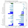
    ETS1 - x axis has the scale, ETS1 is reduced in almost all cell types in people with Long covid at 12 months

    Screen Shot 2024-06-20 at 10.44.25 pm.png
     
  6. SNT Gatchaman

    SNT Gatchaman Senior Member (Voting Rights)

    Messages:
    5,923
    Location:
    Aotearoa New Zealand
    Also highlighting this passage —

    164 is Chronic fatigue syndrome and circulating cytokines: A systematic review (2015, Brain, Behavior, and Immunity) by a certain P.D. White, which stated —

     
  7. SNT Gatchaman

    SNT Gatchaman Senior Member (Voting Rights)

    Messages:
    5,923
    Location:
    Aotearoa New Zealand
    Now published as —

    Sex differences and immune correlates of Long Covid development, symptom persistence, and resolution
    Rebecca E. Hamlin; Shaun M. Pienkos; Leslie Chan; Mikayla A. Stabile; Kassandra Pinedo; Mallika Rao; Philip Grant; Hector Bonilla; Marisa Holubar; Upinder Singh; Karen B. Jacobson; Prasanna Jagannathan; Yvonne Maldonado; Susan P. Holmes; Aruna Subramanian; Catherine A. Blish

    Sex differences have been observed in acute coronavirus disease 2019 (COVID-19) and Long Covid (LC) outcomes, with greater disease severity and mortality during acute infection in males and greater proportions of females developing LC. We hypothesized that sex-specific immune dysregulation contributes to LC pathogenesis.

    To investigate the immunologic underpinnings of LC development and symptom persistence, we performed multiomic analyses on blood samples obtained during acute severe acute respiratory syndrome coronavirus 2 (SARS-CoV-2) infection and 3 and 12 months after infection in a cohort of 45 participants who either developed LC or recovered.

    Several sex-specific immune pathways were associated with LC. Males who would later develop LC exhibited increases in transforming growth factor–β (TGF-β) signaling during acute infection, whereas females who would go on to develop LC had reduced TGFB1 expression. Females who developed LC demonstrated increased expression of XIST, an RNA gene implicated in autoimmunity, during acute infection compared with females who recovered. Many immune features of LC were also conserved across sexes, such as alterations in monocyte phenotype and activation state. Nuclear factor κB (NF-κB) transcription factors were up-regulated in many cell types at acute and convalescent time points. Those with ongoing LC demonstrated reduced ETS1 expression across lymphocyte subsets and elevated intracellular IL-4 in T cell subsets, suggesting that ETS1 alterations may drive aberrantly elevated T helper cell 2–like responses in LC.

    Altogether, this study describes multiple innate and adaptive immune correlates of LC, some of which differ by sex, and offers insights toward the pursuit of tailored therapeutics.

    Link | PDF (Science Translational Medicine) [Open Access]
     
  8. Hutan

    Hutan Moderator Staff Member

    Messages:
    29,697
    Location:
    Aotearoa New Zealand
    This is an interesting paper, especially with the corroboration of the reduced ETS1 finding.

    Google AI said:
    The transcription factor Ets1 is is highly expressed in B cells, particularly in naive B cells, and plays a key role in regulating B cell development and function:

    Expression
    Ets1 levels are high in naive B cells, but rapidly decrease after stimulation. This downregulation occurs within 2–4 hours of stimulation and remains low for at least 12 hours.

    Function
    Ets1 regulates B cell differentiation into plasma cells, and is necessary for maintaining B cells in a quiescent state. Ets1 also regulates antibody production and secretion.

    Autoimmune diseases
    Single-nucleotide polymorphisms (SNPs) in the human ETS1 gene have been linked to a variety of autoimmune diseases, including lupus.

    Blimp1
    Ets1 regulates B cell differentiation by interacting with the transcription factor Blimp1.Ets1 physically interacts with Blimp-1, blocking Blimp-1 DNA binding activity and reducing its ability to repress target genes.

    Target genes
    Ets1 regulates B cell differentiation through direct binding to target genes, including Pax5, which is crucial for maintaining mature B cell identity.

    Ets1 is also expressed in T cells, and mice lacking Ets1 have excess B and T cell activation, leading to enhanced formation of Ab-secreting cells and high titers of autoantibodies. ​

    Ets-1 knockout mice develop increased numbers of IgM-secreting plasma cells. ​
     
    Ron, wigglethemouse, Lindberg and 3 others like this.

Share This Page