Sex differences and immune correlates of Long COVID development, persistence, and resolution, 2024, Hamlin et al.

SNT Gatchaman

Senior Member (Voting Rights)
Staff member
Sex differences and immune correlates of Long COVID development, persistence, and resolution
Rebecca E Hamlin; Shaun M Pienkos; Leslie Chan; Mikayla A Stabile; Kassandra Pinedo; Mallika Rao; Philip Grant; Hector Bonilla; Marisa Holubar; Upinder Singh; Karen B Jacobson; Prasanna Jagannathan; Yvonne Maldonado; Susan P Holmes; Aruna Subramanian; Catherine A Blish

Sex differences have been observed in acute COVID-19 and Long COVID (LC) outcomes, with greater disease severity and mortality during acute infection in males and a greater proportion of females developing LC. We hypothesized that sex-specific immune dysregulation contributes to the pathogenesis of LC.

To investigate the immunologic underpinnings of LC development and persistence, we used single-cell transcriptomics, single-cell proteomics, and plasma proteomics on blood samples obtained during acute SARS-CoV-2 infection and at 3 and 12 months post-infection in a cohort of 45 patients who either developed LC or recovered.

Several sex-specific immune pathways were associated with LC. Specifically, males who would develop LC at 3 months had widespread increases in TGF-β signaling during acute infection in proliferating NK cells. Females who would develop LC demonstrated increased expression of XIST, an RNA gene implicated in autoimmunity, and increased IL1 signaling in monocytes at 12 months post infection.

Several immune features of LC were also conserved across sexes. Both males and females with LC had reduced co-stimulatory signaling from monocytes and broad upregulation of NF-κB transcription factors. In both sexes, those with persistent LC demonstrated increased LAG3, a marker of T cell exhaustion, reduced ETS1 transcription factor expression across lymphocyte subsets, and elevated intracellular IL-4 levels in T cell subsets, suggesting that ETS1 alterations may drive an aberrantly elevated Th2-like response in LC.

Altogether, this study describes multiple innate and adaptive immune correlates of LC, some of which differ by sex, and offers insights toward the pursuit of tailored therapeutics.


Link | PDF (Preprint: BioRxiv) [Open Access]
 
ETS1 (GeneCards)

In both sexes, those with persistent LC demonstrated increased LAG3, a marker of T cell exhaustion, reduced ETS1 transcription factor expression across lymphocyte subsets, and elevated intracellular IL-4 levels in T cell subsets, suggesting that ETS1 alterations may drive an aberrantly elevated Th2-like response in LC.

In summary, this study utilizes multiple modalities to uncover patterns of immune dysregulation that correlate with LC development, persistence, and resolution. Our discovery of sex-specific differences in immune response suggests that future mechanistic and therapeutic studies should consider stratification by sex, as supported by disproportionate impacts of severe acute disease in males and LC in females. We have also found several potential targets for intervention, most notably downregulation of ETS1 transcription factor and increased expression of Th2 cytokines in those with LC.

Corroborated by Blood transcriptomic analyses reveal persistent SARS-CoV-2 RNA and candidate biomarkers in post-COVID-19 condition (2024, The Lancet Microbe) —

Transcript ID ETS1-mRNA
Log2 fold change -0.664
std error (log2) 0.164
Lower confidence limit (log2) -0.986
Upper confidence limit (log2) -0.342
Linear fold change 0.631
Lower confidence limit (linear) 0.505
Upper confidence limit (linear) 0.789
FDR <5% Yes
P value 0.000156
q value 0.012954
Gene.sets probe.ID Growth Factor Signaling NM_005238.3:1305
 
Genecards mentioned a link with Lupus.
Googling ETS1 and Lupus:
The role of the transcription factor Ets1 in lupus and other autoimmune diseases, 2016

One of the important genetic contributions to SLE development is an alteration in the expression of the transcription factor Ets1, which regulates the functional differentiation of lymphocytes. Here we review the genetic, biochemical and immunological studies that have linked low levels of Ets1 to aberrant lymphocyte differentiation and to the pathogenesis of SLE.
 
There's a lot in this study - it's a bit overwhelming in terms of sorting out what is going on and what is worth looking at .

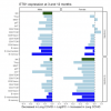
NFKB1, which is a primary component of the canonical NF-κB signaling pathway and induces cell proliferation and immune response programs (151), was found to have higher expression during acute infection in those who will develop LC at 3 months, as well as higher expression at 3 and 12 months in those with LC persisting to 12 months (Fig 7D).

Darker red means a stronger association between higher NFKB1 expression and Long covid at 12 months - for various cell types.
It's notable that there is no blue, which would indicate lower NFKB1 expression.
For males and females combined

Screen Shot 2024-06-20 at 10.25.32 pm.png
 
Also highlighting this passage —

In this cohort, there were notable perturbations in innate and early adaptive immunity during acute infection that correlated with development of LC. Proliferating NK cells in males showed bidirectional TGF-β signaling, which would be expected to reduce NK cell cytotoxicity, as previously observed in severe COVID-19 (161), which may limit the efficacy of the initial antiviral response. Increased TGFβ1 expression was also found to be associated with LC development and persistence across many cell types, including naive and memory T cells, which may lead to reduced T helper 1 (Th1) differentiation (162) and impaired immunity against intracellular pathogens. Conversely, TGF-β signaling contributes to regulatory T cell differentiation, which may be important for avoiding self-reactivity (163). Interestingly, TGF-β has been previously suggested to play a role in myalgic encephalomyelitis/chronic fatigue syndrome (ME/CFS) (164) and was described to be increased in the plasma of males with LC in a pre-print study of a different LC patient cohort (165).

164 is Chronic fatigue syndrome and circulating cytokines: A systematic review (2015, Brain, Behavior, and Immunity) by a certain P.D. White, which stated —

White et al (2015) said:
The observations therefore seem to be consistent with a biologically significant elevation in the cytokine concentration in CFS. The clinical significance of a raised TGF-β is unclear in CFS, but its associations with clinical symptoms, severity and prognosis merits further evaluation.
 
Now published as —

Sex differences and immune correlates of Long Covid development, symptom persistence, and resolution
Rebecca E. Hamlin; Shaun M. Pienkos; Leslie Chan; Mikayla A. Stabile; Kassandra Pinedo; Mallika Rao; Philip Grant; Hector Bonilla; Marisa Holubar; Upinder Singh; Karen B. Jacobson; Prasanna Jagannathan; Yvonne Maldonado; Susan P. Holmes; Aruna Subramanian; Catherine A. Blish

Sex differences have been observed in acute coronavirus disease 2019 (COVID-19) and Long Covid (LC) outcomes, with greater disease severity and mortality during acute infection in males and greater proportions of females developing LC. We hypothesized that sex-specific immune dysregulation contributes to LC pathogenesis.

To investigate the immunologic underpinnings of LC development and symptom persistence, we performed multiomic analyses on blood samples obtained during acute severe acute respiratory syndrome coronavirus 2 (SARS-CoV-2) infection and 3 and 12 months after infection in a cohort of 45 participants who either developed LC or recovered.

Several sex-specific immune pathways were associated with LC. Males who would later develop LC exhibited increases in transforming growth factor–β (TGF-β) signaling during acute infection, whereas females who would go on to develop LC had reduced TGFB1 expression. Females who developed LC demonstrated increased expression of XIST, an RNA gene implicated in autoimmunity, during acute infection compared with females who recovered. Many immune features of LC were also conserved across sexes, such as alterations in monocyte phenotype and activation state. Nuclear factor κB (NF-κB) transcription factors were up-regulated in many cell types at acute and convalescent time points. Those with ongoing LC demonstrated reduced ETS1 expression across lymphocyte subsets and elevated intracellular IL-4 in T cell subsets, suggesting that ETS1 alterations may drive aberrantly elevated T helper cell 2–like responses in LC.

Altogether, this study describes multiple innate and adaptive immune correlates of LC, some of which differ by sex, and offers insights toward the pursuit of tailored therapeutics.

Link | PDF (Science Translational Medicine) [Open Access]
 
This is an interesting paper, especially with the corroboration of the reduced ETS1 finding.

Google AI said:
The transcription factor Ets1 is is highly expressed in B cells, particularly in naive B cells, and plays a key role in regulating B cell development and function:

Expression
Ets1 levels are high in naive B cells, but rapidly decrease after stimulation. This downregulation occurs within 2–4 hours of stimulation and remains low for at least 12 hours.

Function
Ets1 regulates B cell differentiation into plasma cells, and is necessary for maintaining B cells in a quiescent state. Ets1 also regulates antibody production and secretion.

Autoimmune diseases
Single-nucleotide polymorphisms (SNPs) in the human ETS1 gene have been linked to a variety of autoimmune diseases, including lupus.

Blimp1
Ets1 regulates B cell differentiation by interacting with the transcription factor Blimp1.Ets1 physically interacts with Blimp-1, blocking Blimp-1 DNA binding activity and reducing its ability to repress target genes.

Target genes
Ets1 regulates B cell differentiation through direct binding to target genes, including Pax5, which is crucial for maintaining mature B cell identity.

Ets1 is also expressed in T cells, and mice lacking Ets1 have excess B and T cell activation, leading to enhanced formation of Ab-secreting cells and high titers of autoantibodies.​

Ets-1 knockout mice develop increased numbers of IgM-secreting plasma cells.​
 
This is another paper I missed at the time.
It is hard to know how much of it is chance findings in a massive screen for all sorts of things.
TGF beta is of interest but we knew that.

Ets-1 might be relevant but I am beginning to see how important it is to distinguish ETS1 from Ets-1.
That is to say that having a defective ETS1 gene, which might give you lupus, is not at all the same thing as having low production of Ets-1 from what may be a normal ETS1 gene. The upshot I think is that for any study like this if a protein like Ets-1 is altered, or transcription of ETS1 is altered, we want to know whether the gene itself, ETS1, is normal in those cases or has a rogue allele.
 
From The role of the transcription factor Ets1 in lupus and other autoimmune diseases 2016:

As early as 2000, polymorphisms in the 3′ UTR of the human ETS1 gene were associated with particular clinical phenotypes of lupus. As described in more detail in the sections below, a few years later, Ets1−/− mice were discovered to develop a lupus-like autoimmune disease, supporting a role for Ets1 in regulating immune tolerance to self-antigens. More recently, three independent genome-wide association studies in Chinese and Korean populations have identified genetic variants in and around ETS1 gene as increasing the lupus risk. These initial genome-wide association studies were later replicated in independent populations of Chinese and Malaysian origin.

As indicated in Table 1, the SNPs associated with lupus in these particular studies all map near the 3′ end of the gene, either in the final intron, in the 3′UTR or downstream of the gene. Exome sequencing in healthy donors and lupus patients has identified a single nucleotide polymorphism (SNP rs34846069) in the final exon of the gene that is associated with lupus, although this SNP does not change the encoded amino acid (Asp440→Asp). This SNP may be in linkage disequilibrium with other genetic changes that promote lupus.

In addition to lupus, SNPs in or near the ETS1 gene have also been identified as susceptibility alleles in many other autoimmune and inflammatory diseases (Table 1), including rheumatoid arthritis, psoriasis, multiple sclerosis, ankylosing spondylitis, uveitis, allergy, atopic dermatitis, and celiac disease.
 
In addition to lupus, SNPs in or near the ETS1 gene have also been identified as susceptibility alleles in many other autoimmune and inflammatory diseases (Table 1), including rheumatoid arthritis, psoriasis, multiple sclerosis, ankylosing spondylitis, uveitis, allergy, atopic dermatitis, and celiac disease.

This is very interesting, if true. Lupus, RA and MS are all antibody-related diseases. Psoriasis, AS and unveitis are not. Complement defects predispose to lupus almost certainly by making it easier to make autoantibody. Interferons may do the same. TLR7 mutations probably impact B cell signalling.

But it looks as if ETS1 can screw up the immune system on a wider basis and meddle with non-antibody diseases too. Although I think immunologists are badly mistaken in thinking that T cell responses control B cell responses (and thus autoimmunity) one thing that is true of all these diseases is that T cell population expansions are probably involved - because there are different sorts of T cells for all sorts of up and down functions - permissions and prohibitions perhaps.

A motivation behind our Qeios piece was that Jo C and I have both had a sense that ME/CFS is anti-inflammatory or maybe anti-autoimmune in some peculiar way. The paper was an attempt to make that consistent with the female predominance.

The data are not all in yet but I am pretty sure that this is the area where we are suddenly going to understand what is going on. Interferons look relevant - whether Rob Phair and @jnmaciuch are right with type I or we are with type 2. CD24, cell maturation and forking of metabolic pathway usage seem relevant. Some sort of trade off between 'vigour' of certain T cell populations and 'overkill', as in Ank Spond seems relevant. ETS1 might be in the middle of all these things. But there is some way in which ME/CFS is backwards from all the others. That is not surprising because each of these diseases is different in some idiosyncratic way. ME/CFS is just awkward because it sends an 'invisible' pathway into overdrive. Maybe a prohibition pathway.


And again, I am thinking that we must not confuse defects in genome with low levels of gene transcription. Complement is a good lesson there. Complement gene defects confer risk of lupus by making it easier to make autoantibody but, through a quite different mechanism they also facilitate the pathology of immune complex damage once autoantibody has been made.
 
Back
Top Bottom