[68Ga]FAPI PET/CT reveals increased pulmonary fibroblast activation protein expression in long COVID patients after ICU discharge, 2025, Leer et al

forestglip

Administrator
Staff member
[68Ga]FAPI PET/CT reveals increased pulmonary fibroblast activation protein expression in long COVID patients after ICU discharge

B. van Leer, M. Londema, Ö. Kasalak, J. H. van Snick, M. L. Duiverman, J. C. Kuijvenhoven, M. D. de Kruif, D. E. Oprea-Lager, K. Pabst, M. E. Hellemons, H. H. Boersma, M. Prokop, M. W. Nijsten, A. W. J. M. Glaudemans, J. Pillay, R. H. J. A. Slart on behalf of the COVID-CLIMATE consortium

[Line breaks added]


Purpose
Post-acute sequelae of COVID-19 (PASC) has emerged as a major healthcare problem. A comprehensive mechanism of disease remains to be elucidated. In this study we aimed to explore pulmonary and muscle fibroblast activation protein (FAP) activity in former critical COVID-19 patients with persistent dyspnea, using [68Ga]FAPI-46 PET/CT.

Methods
In this single center prospective observational study we included former critical COVID-19 patients reporting complaints of dyspnea > 3 months after hospital discharge. A [68Ga]FAPI PET/CT scan was performed including a high-resolution CT scan, lung function test, EQ-5D questionnaire, 6 min walking test and inflammatory markers.

Age and sex-matched subjects, without pulmonary pathology, served as controls. The [68Ga]FAPI uptake was corrected for lean body mass and the target-to-background ratio (TBR) was calculated.

Results
Eighteen PASC patients and 15 controls (median age 59 and 63 years and BMI of 34.6 and 25.2 kg/m2) were included. The interval between hospital discharge and study visit was 30 months.

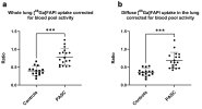
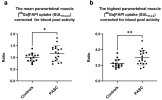
Increased pulmonary FAP expression was observed in PASC, (TBR 0.79 ± 0.23) compared to controls (TBR 0.40 ± 0.13, P < 0.001). Increased FAP expression was also observed in the paravertebral muscles (PASC: TBR 1.17 and controls TBR 1.00, P = 0.03).

Forced expiratory volume and forced vital capacity showed moderate negative correlation with the pulmonary TBR, while the percentage of ground glass opacities showed a moderate positive correlation.

Conclusion
[68Ga]FAPI PET/CT demonstrated elevated FAP expression in PASC. These findings provide insight into possible pathophysiological mechanisms of PASC and a potential new diagnostic modality.

Link | PDF (European Journal of Nuclear Medicine and Molecular Imaging) [Open Access]
 
Molecular imaging with 68 Ga-fibroblast activation protein inhibitor ([68 Ga]FAPI) positron emission tomography (PET) allows for whole body assessment of in vivo fibroblast activation. [68 Ga]FAPI binds to fibroblast activation protein (FAP) which is expressed on activated fibroblasts. Our aim was to explore pulmonary FAP activity in former critical COVID-19 patients, with persistent complaints of dyspnea. We hypothesised that PASC patients show a higher pulmonary [68 Ga]FAPI uptake compared to controls.

Overall, lung function parameters were within normal limits in most patients (median percentage of predicted diffuse capacity for carbon monoxide (DCLO) was 79% (66 to 89), forced expiratory volume (FEV1) 90% (81 to 104) and forced volume capacity (FVC) 86% (74 to 92)). Median walking distance during the 6MWT was 393 meters (228 to 482).

Increased pulmonary [68 Ga]FAPI uptake (TBRwl ) was observed in the PASC patients (mean TBR: 0.79 ± 0.23) compared to controls (mean TBR 0.40 ± 0.13, P < 0.001).

Paravertebral muscle uptake was visually observed and post-hoc analyses showed significant difference between PASC and controls for the mean and maximal [68 Ga]FAPI uptake corrected for blood pool activity (TBR PASC: 1.17 (± 0.33) and controls: 1.00 (± 0.17), P= 0.03 and TBR PASC: 2.9 (± 0.92) and controls: 2.2 (± 0.54), P= 0.004. The increase in fatigue following the 6MWT correlates with both the mean and maximal [68 Ga]FAPI muscle uptake (r =0.70, P = 0.002 and r= 0.65, P= 0.005). Whereas the increase of dyspnea after the 6 MWT, the EQ-index and the EQ-VATS did not correlate.

More than 2 years after hospital discharge, markedly increased pulmonary [68 Ga]FAPI uptake was observed in PASC compared to controls. […] In addition, we found increased FAP expression in muscle tissue of PASC patients.

These findings were confirmed in a small prospective study including 6 PASC patients and 9 controls]. However, this study only included patients with persistent CT abnormalities, which are frequently absent in PASC. In this study, we found increased long-term pulmonary FAP expression, only moderately correlated to the presence of ground glass opacities.

To our knowledge this is the first time that increased muscle FAP expression in PASC patients has been shown. Skeletal muscle involvement in the presence of exertion related PASC symptoms has been described [18].

[18] is Muscle abnormalities worsen after post-exertional malaise in long COVID (2024)
 
So, while aiming to evaluate activated fibroblasts in lung, they noted uptake in the paravertebral muscles that happened to be in the imaging range.

Screenshot 2025-06-25 at 9.37.49 AM copy.jpg
Screenshot 2025-06-25 at 9.38.19 AM copy.jpgScreenshot 2025-06-25 at 9.38.33 AM copy.jpg

This is something Rob Wüst and Anouk Slaghekke will be interested in given their poster presentation on collagen IV deposition in muscle capillary basement membranes. (Fibroblasts might also be responsible for their previously observed amyloid deposits, or may become activated by amyloid produced by something else and deposited.)

[68 Gd]FAPI could be applied to skeletal muscle. See eg Diagnostic and evaluative efficiency of 68Ga-FAPI-04 in skeletal muscle injury (2024)
 
So we have an independent replication of this finding, and genetic evidence that an isoform of BTN2A1 implicated in MECFS is highly expressed in fibroblasts*. This area seems like it could be well worth digging into, as @DMissa is planning to.

[*Edit for clarity: high fibroblast expression of this isoform is in general, not in MECFS specifically.]
 
Last edited:
So we have an independent replication of this finding, and genetic evidence that an isoform of BTN2A1 implicated in MECFS is highly expressed in fibroblasts*.
Sorry what's the common thread here? What's the link between FAP and BTN2A1?
 
Sorry what's the common thread here? What's the link between FAP and BTN2A1?
Ive possibly got mixed up! I thought FAP showed up in fibroblasts that were 'activated' because of immune activity in general but upon Googling it seems like it's specifically involved in tumours and the extracellar matrix?

I suppose I was thinking FAP showing up in two studies might be a sign that something strange may be afoot
with fibroblasts, signalling wise. Something which could possibly be linked to BTN2A1 associated immune signalling abnormalities perhaps. But it's very likely I've gotten something completely wrong!

It was partly this passage from Wikipedia that got me thinking along these lines.
Fibroblasts from different anatomical sites in the body express many genes that code for immune mediators and proteins.[16] These mediators of immune response enable the cellular communication with hematopoietic immune cells.[17] The immune activity of non-hematopoietic cells, such as fibroblasts, is referred to as "structural immunity".[16][18] In order to facilitate a fast response to immunological challenges, fibroblasts encode crucial aspects of the structural cell immune response in the epigenome.

I'm out of my depth here with my dimly remembered GCSE biology qualification and bits and bobs I've gleaned from reading studies on here, as I'm sure you can tell.
 
Last edited:
I thought FAP showed up in fibroblasts that were 'activated' because of immune activity in general but upon Googling it seems like it's specifically involved in tumours and the extracellar matrix?
Perhaps the opposite ? The innate immune system might perhaps be activated by suspicious activity in the fibroblasts which persists after an infection.
 
might be a sign that something strange may be afoot
with fibroblasts
For ME/CFS the way I look at fibroblasts is a way to test whether a suspected problem is systemic or not, using a tissue that can be collected and turned into a primary cell line and bunged into the freezer for whenever questions arise relatively easily. I am not sure how an issue intrinsic to skin cells would mediate ME/CFS. Probably shouldn't tie our brains in knots over it imo

If there is an observation made in fibroblasts that stands out as interesting, the next step would probably be to think about what other tissues/cells of plausible relevance to an ME/CFS disease mechanism could be affected by a similar issue in a way that could contribute to the illness in specific terms

for example if someone saw something happening with IFN-β in fibros that could be grist for the mill because of applicability to systems we think are likely to be relevant
 
Last edited:
For ME/CFS the way I look at fibroblasts is a way to test whether a suspected problem is systemic or not, using a tissue that can be collected and turned into a primary cell line and bunged into the freezer for whenever questions arise relatively easily. I am not sure how an issue intrinsic to skin cells would mediate ME/CFS. Probably shouldn't tie our brains in knots over it imo

If there is an observation made in fibroblasts that stands out as interesting, the next step would probably be to think about what other tissues/cells of plausible relevance to an ME/CFS disease mechanism could be affected by a similar issue in a way that could contribute to the illness in specific terms

for example if someone saw something happening with IFN-β in fibros that could be grist for the mill because of applicability to systems we think are likely to be relevant
Thanks that's informative.

Even if it's well downstream of any mechanism, I do wonder about this FAP marker and whether we'd see it in MECFS too. If we do it could be useful for cohort selection and maybe even as a marker for clinical trials if it corresponded to severity (I can't remember if they mention that in these LC studies).

Edit: on second thought these scans are so expensive their use would inflate trial budgets through the roof. Perhaps it would be useful for diagnosis though.
 
Last edited:
Back
Top Bottom