Nightsong

Senior Member (Voting Rights)
Abstract:
We proposed that cerebrospinal fluid would provide objective evidence for disrupted brain metabolism in myalgic encephalomyelitis/chronic fatigue syndroome (ME/CFS). The concept of postexertional malaise (PEM) with disabling symptom exacerbation after limited exertion that does not respond to rest is a diagnostic criterion for ME/CFS. We proposed that submaximal exercise provocation would cause additional metabolic perturbations. The metabolomic and lipidomic constituents of cerebrospinal fluid from separate nonexercise and postexercise cohorts of ME/CFS and sedentary control subjects were contrasted using targeted mass spectrometry (Biocrates) and frequentist multivariate general linear regression analysis with diagnosis, exercise, gender, age and body mass index as independent variables.

ME/CFS diagnosis was associated with elevated serine but reduced 5-methyltetrahydrofolate (5MTHF). One carbon pathways were disrupted. Methylation of glycine led to elevated sarcosine but further methylation to dimethylglycine and choline was decreased. Creatine and purine intermediates were elevated. Transaconitate from the tricarboxylic acid cycle was elevated in ME/CFS along with essential aromatic amino acids, lysine, purine, pyrimidine and microbiome metabolites. Serine is a precursor of phospholipids and sphingomyelins that were also elevated in ME/CFS. Exercise led to consumption of lipids in ME/CFS and controls while metabolites were consumed in ME/CFS but generated in controls.

The findings differ from prior hypometabolic findings in ME/CFS plasma. The novel findings generate new hypotheses regarding serine-folate-glycine one carbon and serine-phospholipid metabolism, elevation of end products of catabolic pathways, shifts in folate, thiamine and other vitamins with exercise, and changes in sphingomyelins that may indicate myelin and white matter dysfunction in ME/CFS.

Link | PDF (Nature Scientific Reports, March 2025, open access)
 
Serine had interest maybe three decades ago, transaconitate sounds like something I might have discussed two and a half decades ago; purines have always been high with me, leading to a wrong diagnosis of gout. I am not sure what they mean by metabolites consumed in exercise, guess I will have to read the paper. Take my mind off the cyclone that may hit my area in a day or three. [cyclone discussion moved here]

If we have broken energy metabolism then the cell finds other pathways and metabolites. However most tissues might have trouble consuming lactate, which means the liver has to consume it all, and it does so slowly.
 
Last edited by a moderator:
Not to sound pedantic, but a typo in the first sentence of a paper published in Nature Sci Reports? "syndroome"

Not to sound pedantic, but he uses “we” in the abstract for a single author paper.

Also getting red flags for a single author paper with so much data
 
Isn’t ‘we’ even for a single author research paper standard practice. Had it been an opinion piece that would have been different.
 
Not to sound pedantic, but a typo in the first sentence of a paper published in Nature Sci Reports? "syndroome"

Scientific Reports, despite being published by Nature [Publishing Group], is not a quality journal, but this fact isn't really appreciated outside of academia. They accept a large percentage of submitted papers, very often publish trash, and the journal has been implicated in numerous scientific fraud cases.
 
This paper says:
Follow-up data analysis including Bayesian statistics are reported separately124.
124 is that other paper. It does appear that the other paper has other results not included in this one, and the wording is changed throughout the paper.

I haven't read either of these in detail, but just as a rough measure, there are 1782 words in this paper's Results section, and 2818 in the "Exertional Exhaustion" paper's Results.

Edit: I think all of the reported results might be based on different methodologies in the other paper.
 
Last edited:
I noticed that several of the citations don't seem to make sense. 3 of the 4 citations in this sentence about metabolites seem to be for papers that are unrelated to metabolites:
Serine had the most correlations[34], followed by 7-methylguanosine[13], creatine[13], 1-methyladenosine[10], and indoleacrylic acid[8].
8. Bae, J. & Lin, J. M. S. Healthcare utilization in myalgic encephalomyelitis/chronic fatigue syndrome (ME/CFS): Analysis of US ambulatory healthcare data, 2000–2009. Front. Pediatr. 7, 185 (2019).

10. Ghali, A. et al. Factors influencing the prognosis of patients with myalgic encephalomyelitis/chronic fatigue syndrome. Diagnostics (Basel) 12(10), 2540 (2022).

13. Jason, L. A., Evans, M., So, S., Scott, J. & Brown, A. Problems in defining post-exertional malaise. J. Prev. Interv. Commun. 43(1), 20–31 (2015).

34. Vance, J. E. Phospholipid synthesis and transport in mammalian cells. Traffic 16(1), 1–18 (2015).
 
Last edited:
The study looked at CSF from two different cohorts: ME/CFS and controls who had not done exercise, and a different group that had just done 2 CPETs.
Two cohorts were studied22,23,24,25. The first day of the 2 protocols was considered an adjustment period, and included the patient’s history and physical, blood work, and baseline studies. The nonexercise cohort rested before having lumbar puncture and did not have exercise. The postexercise cohort had magnetic resonance imaging (MRI), submaximal bicycle exercise stress testing, and serial assessments of postural tachycardia. They rested overnight, then had their second identical stress test, MRI and postexercise lumbar puncture. Subjects cycled at 70% of predicted heart rate (pHR = 220-Age) for 25 min then increased to 85% pHR. Lumbar puncture was performed 1 to 5 h after exercise.

The largest difference was increased level of serine in cerebrospinal fluid in ME/CFS, and this metabolite also had the most correlations to symptom scores in ME/CFS, so this seems like a somewhat strong finding.
ME/CFS had higher levels of serine (Hedges’ g = 0.962), five sphingomyelins and five other phospholipids than SC with elevated 1-methylhistidine (g = -0.531) in SC [sedentary controls].
Analyte abundance was correlated with questionnaire items to find associations between potential markers of disease and disease manifestations. The nonparametric Spearman’s correlation matrix was inspected and the quadrant of analytes vs. items excised. Features were sorted to find the metabolites and lipids with the most relationships with questionnaire items. Significance of this process was defined by |R| > 0.3 (p < 0.015). Three general patterns were found (Fig. 5, SOM Table S4). Serine had the most correlations[34], followed by 7-methylguanosine[13], creatine[13], 1-methyladenosine[10], and indoleacrylic acid[8]. This set was correlated with Multidimensional Fatigue Index Reduced Activity (11 analytes), Physical Fatigue[8] and sum[8], Mystery[9], Permanance[7] and CPSS[7]. Elevated serine and other metabolites in ME/CFS > SC were correlated with fatigue which was consistent with the diagnostic criteria of ME/CFS.

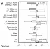
It looks like serine was higher in ME/CFS than in controls in both cohorts, even when splitting by sex (though only significantly in 2/4 comparisons). Fig 2A:
1786931289327.webp
 
Last edited:
I'm not sure if the study is measuring both D-serine and L-serine at once, or if it's one or the other. According to Wikipedia, D-serine has a neuron-related role:
D-Serine, synthesized in neurons by serine racemase from L-serine (its enantiomer), serves as a neuromodulator by coactivating NMDA receptors, making them able to open if they then also bind glutamate. D-serine is a potent agonist at the glycine site (NR1) of canonical diheteromeric NMDA receptors. For the receptor to open, glutamate and either glycine or D-serine must bind to it; in addition a pore blocker must not be bound (e.g. Mg2+ or Zn2+).[14] Some research has shown that D-serine is a more potent agonist at the NMDAR glycine site than glycine itself.[15][16] However, D-serine has been shown to work as an antagonist/inverse co-agonist of t-NMDA receptors through the glycine binding site on the GluN3 subunit.[17][18]
 
Just quickly searching for what other diseases might have increased serine. Possibly Alzheimer's disease and bipolar disorder.

D-serine levels in Alzheimer’s disease: implications for novel biomarker development (Madeira et al., 2015)
Finally, we assessed d-serine levels in the cerebrospinal fluid (CSF) of 21 patients with a diagnosis of probable AD [Alzheimer's disease], as compared with patients with normal pressure hydrocephalus (n=9), major depression (n=9) and healthy controls (n=10), and results were contrasted with CSF amyloid-β/tau AD biomarkers. d-serine levels were higher in the hippocampus and parietal cortex of AD patients than in control subjects. Levels of both d-serine and serine racemase, the enzyme responsible for d-serine production, were elevated in experimental models of AD. Significantly, d-serine levels were higher in the CSF of probable AD patients than in non-cognitively impaired subject groups.

CSF d-serine concentrations are similar in Alzheimer's disease, other dementias, and elderly controls (Biemans et al., Neurobiology of Aging, 2016)
In contrast to previous findings, in our study CSF d-serine levels were only slightly increased in AD patients compared with controls. CSF d-serine in AD did not differ from other dementias and was also not correlated to mini-mental state examination–scores.

Cerebrospinal Fluid and Serum d-Serine Levels in Patients with Alzheimer’s Disease: A Systematic Review and Meta-Analysis (Chang et al., J. Clin. Med., 2020)
Seven trials with more than 1186 participants were included in this meta-analysis. d-serine levels in patients with AD were significantly higher than those in controls (SMD = 0.679, 95% CI = 0.335 to 1.022, p < 0.001). Subgroup analyses showed that the AD group had significantly higher d-serine levels in serum and CSF compared with the control group (SMD = 0.566 (serum) and 1.008 (CSF); 95% CI = 0.183 to 0.948 (serum) and 0.168 to 1.849 (CSF)).

Markers of glutamate signaling in cerebrospinal fluid and serum from patients with bipolar disorder and healthy controls (Pålsson et al., 2015)
A total of 215 patients with bipolar disorder and 112 healthy controls from the Swedish St. Göran bipolar project were included in this study. [...] Serum levels of glutamine, glycine and d-serine were significantly higher whereas l-serine levels were lower in patients with bipolar disorder as compared to controls.
 
Back
Top Bottom