Genetics: Chromosome 6 BTN2A2 and BTN3A3 (BTN2A1)

BTN2A1_tissue_expression.webp
Figures for the different BTN2A1 isoforms and their tissue-specific transcript expression, as promised. As a reminder, most of the ME/CFS-associated mutations fell in the PRY-SPRY domain, which is only present in 2/4 isoforms (the two on the left in the figure).

I forgot I can't upload a markdown document, but here's the code:
Code:
---
title: "Tissue-specific expression analysis of BTN isoforms"
output: html_document
date: "2026-08-29"
---

```{r setup, include=FALSE}
knitr::opts_chunk$set(echo = TRUE)

# Set working directory
workDir <- "~/Documents/coding"

# Load packages
library(jsonlite)
library(dplyr)
library(ggplot2)
```

BTN2A1 has several isoforms with different protein domains. Per the [AstraZeneca](https://azphewas.com/QVView/6319c068-fd59-46d8-85ee-82d82482eb14/BTN2A1/8e436248-c6c7-4c3c-9520-6a03495b72af/f5e4d803-b578-4371-9c76-b43baf74d772/db9a8d3d-b11a-4b5d-a129-4327844bcfb1/binary/EUR) rare mutation analysis, most ME/CFS-associated mutations were in the cytosolic PRY-SPRY domain.

Per [UniProt](https://www.uniprot.org/uniprotkb/Q7KYR7/entry), we know of 4 protein isoforms of BTN2A1. From the "Genome annotation databases" section, I manually pulled the associated transcript IDs (e.g. ENST00000312541.10) for all listed isoforms (only the ones matching BTN2A1 GENCODE ID ENSG00000112763.18, since ENSG00000292170.1 is not used in GTEx).

Clicking on the transcript ID link takes you to the Ensembl entry, where I found information on whether the isoform contained an SPRY domain via the "Transcript function" button. I compiled this information manually into a spreadsheet.

```{r}

# Load table for Uniprot transcripts and isoforms of BTN2A1
BTN_df <- read.csv(file.path(workDir,
                             "BTN2A1_isoform_table.csv"))
```

To test the hypothesis that certain protein isoforms with or without this protein domain had different expression levels in the cerebellum and/or immune cells, I pulled the median transcript expression values from the GTEx API using the call:

```{bash}
curl -X 'GET' \
  'https://gtexportal.org/api/v2/expression/medianTranscriptExpression?gencodeId=ENSG00000112763.17&datasetId=gtex_v10&tissueSiteDetailId=Adrenal_Gland&tissueSiteDetailId=Brain_Amygdala&tissueSiteDetailId=Brain_Anterior_cingulate_cortex_BA24&tissueSiteDetailId=Brain_Caudate_basal_ganglia&tissueSiteDetailId=Brain_Cerebellar_Hemisphere&tissueSiteDetailId=Brain_Cerebellum&tissueSiteDetailId=Brain_Cortex&tissueSiteDetailId=Brain_Frontal_Cortex_BA9&tissueSiteDetailId=Brain_Hippocampus&tissueSiteDetailId=Brain_Hypothalamus&tissueSiteDetailId=Brain_Nucleus_accumbens_basal_ganglia&tissueSiteDetailId=Brain_Putamen_basal_ganglia&tissueSiteDetailId=Brain_Spinal_cord_cervical_c-1&tissueSiteDetailId=Brain_Substantia_nigra&tissueSiteDetailId=Breast_Mammary_Tissue&tissueSiteDetailId=Cells_Cultured_fibroblasts&tissueSiteDetailId=Cells_EBV-transformed_lymphocytes&tissueSiteDetailId=Colon_Sigmoid&tissueSiteDetailId=Colon_Transverse&tissueSiteDetailId=Liver&tissueSiteDetailId=Lung&tissueSiteDetailId=Minor_Salivary_Gland&tissueSiteDetailId=Muscle_Skeletal&tissueSiteDetailId=Nerve_Tibial&tissueSiteDetailId=Pancreas&tissueSiteDetailId=Pituitary&tissueSiteDetailId=Skin_Not_Sun_Exposed_Suprapubic&tissueSiteDetailId=Skin_Sun_Exposed_Lower_leg&tissueSiteDetailId=Small_Intestine_Terminal_Ileum&tissueSiteDetailId=Spleen&tissueSiteDetailId=Stomach&tissueSiteDetailId=Thyroid&tissueSiteDetailId=Whole_Blood&tissueSiteDetailId=Adipose_Subcutaneous&tissueSiteDetailId=Adipose_Visceral_Omentum&tissueSiteDetailId=Artery_Aorta&tissueSiteDetailId=Artery_Coronary&tissueSiteDetailId=Artery_Tibial&tissueSiteDetailId=Bladder&tissueSiteDetailId=Cervix_Ectocervix&tissueSiteDetailId=Cervix_Endocervix&tissueSiteDetailId=Esophagus_Gastroesophageal_Junction&tissueSiteDetailId=Esophagus_Mucosa&tissueSiteDetailId=Esophagus_Muscularis&tissueSiteDetailId=Fallopian_Tube&tissueSiteDetailId=Heart_Atrial_Appendage&tissueSiteDetailId=Heart_Left_Ventricle&tissueSiteDetailId=Kidney_Cortex&tissueSiteDetailId=Kidney_Medulla&tissueSiteDetailId=Ovary&tissueSiteDetailId=Prostate&tissueSiteDetailId=Testis&tissueSiteDetailId=Uterus&tissueSiteDetailId=Vagina&page=0&itemsPerPage=100000' \
  -H 'accept: application/json'
```

```{r}

# Import transcript counts obtained from API call
counts <- fromJSON(file.path(workDir,
                             "response_1788031490143.json"))$data

```

Note that I am making an assumption that detection of the alternatively spliced transcript correlates to actual protein expression.

This call pulls all available transcript counts for the gene, so I subset to transcripts that had information in Uniprot:

```{r}

# Keep only counts for transcript IDs in table
counts <- inner_join(BTN_df,
                     counts,
                     by = "transcriptId")

```

And now I plot the results:

```{r}

# Some additional formatting for plotting:
counts <- counts %>%
  mutate(SPRY_domain = if_else(SPRY_domain, "Contains SPRY domain", "No SPRY domain"),
         SPRYId = paste(SPRY_domain, uniprotId, sep = " | "))

# Load color scheme for tissues (from https://github.com/broadinstitute/gtex-v8/blob/master/data/gtex_colors.txt)
color_df <- read.delim(file.path(workDir,
                               "gtex_colors.txt"))

# Create color scale vector
color_scale <- c(color_df$color_hex) %>% setNames(color_df$tissue_id)

# Plot
ggplot(counts,
       aes(y = tissueSiteDetailId,
           x = median)) +
  geom_col(aes(fill = tissueSiteDetailId)) +
  scale_fill_manual(values = color_scale) +
  facet_wrap(vars(SPRYId, transcriptId), nrow = 1) +
  theme_bw() +
  theme(axis.text.x = element_text(angle = 90, hjust = 1)) +
  guides(fill = "none")
```

I plotted all available organs, specifically paying attention to the cerebellum and immune cell "tissues" (Whole Blood, Spleen, EBV-transformed lymphoblasts, cultured fibroblasts). While the two SPRY-containing isoforms (Q7KYR7-2 and Q7KYR7-5) had highest levels in the cerebellum, each was roughly matched by at least one immune tissue (fibroblasts and spleen, respectively). High expression in the arterial tissues matching cerebellar expression was also notable.

Overall, there is not a strong and clear indication that the BTN2A1 isoforms containing regions with ME/CFS-associated mutations are much more strongly expressed in brain vs. immune cells. Though analysis is limited by lack of resolution on specific immune cell types.

And the referenced files are attached
 

Attachments

Last edited:
While the two SPRY-containing isoforms (Q7KYR7-2 and Q7KYR7-5) had highest levels in the cerebellum, each was roughly matched by at least one immune tissue (fibroblasts and spleen, respectively). High expression in the arterial tissues matching cerebellar expression was also notable.
This is really interesting. Makes me think again about the idea of BTNs doing something in both brain and immune tissues in MECFS, rather than one or the other.
 
This is really interesting. Makes me think again about the idea of BTNs doing something in both brain and immune tissues in MECFS, rather than one or the other.
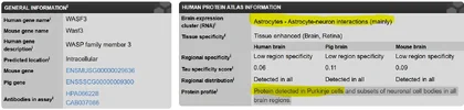
And also I should reiterate, this is bulk tissue expression. Meaning that theoretically some of the signal from brain tissues could be microglia/astrocytes/etc. Though if that were the case here, there’d have to be an explanation for why microglia in the cerebellum are different than any other part of the brain.

I know that some studies have found variation across a bunch of different genes when sampling microglia from different brain regions but I don’t remember seeing much about a gene with such a strong difference like this.
 
Apparently the cerebellum does have a unique glial cell type that integrates "noxious" stimuli....
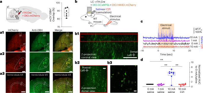
The cerebellum is activated by noxious stimuli and pathological pain but its role in noxious information processing remains unknown. Here, we show that in mice, cutaneous noxious electrical stimuli induced noradrenaline (NA) release from locus coeruleus (LC) terminals in the cerebellar cortex. Bergmann glia (BG) accumulated these LC–NA signals by increasing intracellular calcium in an integrative manner (‘flares’). BG flares were also elicited in response to an intraplantar capsaicin injection. Chemogenetic inactivation of LC terminals or BG in the cerebellar cortex or BG-specific knockdown of α1-adrenergic receptors suppressed BG flares, reduced nocifensive licking and had analgesic effects in nerve injury-induced chronic neuropathic pain. Moreover, chemogenetic activation of BG or an intraplantar capsaicin injection reduced Purkinje cell firing, which may disinhibit the output activity of the deep cerebellar nuclei. These results suggest a role for BG in computing noxious information from the LC and in modulating pain-related behaviors by regulating cerebellar output.
 
Just for general information

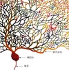
Human Purkinje cells ( cerebellum)can host up to 200,000 dendritic spines (the tiny contact points that receive chemical signals), allowing a single cell to connect with hundreds of thousands of other neurons.Purkinje neurons have the most extensive dendritic branching of all neurones.
 

Attachments

  • 20210412-purkinje-neuron-JA.webp
    20210412-purkinje-neuron-JA.webp
    133.8 KB · Views: 7
Do you mean between cerebellum and other brain tissues? And if so could you expand on it?

Yes, sorry, between cerebellum and other brain areas. The tissue is dominated by Purkinje cells. These have vast trees of ramifying dendrites in highly stereotyped bunches, branching on branching. In contrast, most large neurons have a main linear dendrite with side branches or sometimes two opposing main dendrites. It is worth looking up pictures on the net.

Purkinje cells presumably integrate incoming information in a completely different way. It is known that they have two styles of input. There are inputs to each of the tops of the tiniest branches but also input coming up from below on to the 'stems' that control the bunch integration.
 
Apparently the cerebellum does have a unique glial cell type that integrates "noxious" stimuli.

Very intriguing.

From Wikipedia:
Bergmann glia (also known as radial epithelial cells, Golgi epithelial cells, or radial astrocytes) are unipolar astrocytes derived from radial glia that are intimately associated with Purkinje cells in the cerebellum. Since Bergmann glia appear to persist in the cerebellum, and perform many of the roles characteristic of astrocytes, they have also been called "specialized astrocytes. Bergmann glia have multiple radial processes that extend across the molecular layer of the cerebellar cortex and terminate at the pial surface as a bulbous endfoot. Bergmann glial cells assist with the migration of granule cells, guiding the small neurons from the external granular layer down to the internal granular layer along their extensive radial processes. Besides their role in early development of the cerebellum, Bergmann glia are also required for synaptic pruning. Following Purkinje cell death induced by CNS injury, Bergmann glia undergo extensive proliferative changes so as to replace lost or damaged tissue in a process known as gliosis.

It seems that these are a type of astrocyte (rather than immune microglia) related to neuron progenitor or stem cells. Structurally they are much more like neurons than anything else and perhaps should be seen as 'housekeeper metaneurons' that do not process electrical signals quite the way neurons do but are nevertheless directly involved in signalling via both immediate and long term modulation of synaptic firing.

The seem to be involved, like CA10, both in noxious stimuli and synaptic pruning.
To me this is very significant. Fifty years ago we were essentially taught that the cerebellum was a vast number crunching station for co-ordinating motor output routines. There was no suggestion that it was involved in sensory 'value' - i.e. noxious value. There have been some other suggestions that cerebellum might be relevant and this makes me think we should take it very seriously. I note that the noxious stimuli seem to be coming in to cerebellum from axons from nucleus caeruleus, which is also involved in sleep and was already on the radar.
 
It looks as if Bergmann glia may be involved in slow modulation of the effects of ON and NC inputs that Marr claimed modulated the top inputs via 'mossy fibres'. It makes me think of a harp analogy. The mossy fibre inputs are like the harpist's fingers flitting over the strings (Purkinje dendrites). The ON and NC inputs are like the pedals that the harpist uses to change key by altering the tension in certain strings. The Bergmann glia are then like the tuning hammers the harpist can use to control tension in the longer term.

Such a system could provide a means of long term distortion of nociceptor pathways that would make much more sense than the backwards 'predictive coding' idea.
 
Very intriguing.

From Wikipedia:
Bergmann glia (also known as radial epithelial cells, Golgi epithelial cells, or radial astrocytes) are unipolar astrocytes derived from radial glia that are intimately associated with Purkinje cells in the cerebellum. Since Bergmann glia appear to persist in the cerebellum, and perform many of the roles characteristic of astrocytes, they have also been called "specialized astrocytes. Bergmann glia have multiple radial processes that extend across the molecular layer of the cerebellar cortex and terminate at the pial surface as a bulbous endfoot. Bergmann glial cells assist with the migration of granule cells, guiding the small neurons from the external granular layer down to the internal granular layer along their extensive radial processes. Besides their role in early development of the cerebellum, Bergmann glia are also required for synaptic pruning. Following Purkinje cell death induced by CNS injury, Bergmann glia undergo extensive proliferative changes so as to replace lost or damaged tissue in a process known as gliosis.

It seems that these are a type of astrocyte (rather than immune microglia) related to neuron progenitor or stem cells. Structurally they are much more like neurons than anything else and perhaps should be seen as 'housekeeper metaneurons' that do not process electrical signals quite the way neurons do but are nevertheless directly involved in signalling via both immediate and long term modulation of synaptic firing.

The seem to be involved, like CA10, both in noxious stimuli and synaptic pruning.
To me this is very significant. Fifty years ago we were essentially taught that the cerebellum was a vast number crunching station for co-ordinating motor output routines. There was no suggestion that it was involved in sensory 'value' - i.e. noxious value. There have been some other suggestions that cerebellum might be relevant and this makes me think we should take it very seriously. I note that the noxious stimuli seem to be coming in to cerebellum from axons from nucleus caeruleus, which is also involved in sleep and was already on the radar.
This seems like a very plausible location for much of the symptom causing pathology of MECFS.
 
Back
Top Bottom