Genetics: Chromosome 6 BTN2A2 and BTN3A3 (BTN2A1)

According to the AstraZeneca PHEWAS, the variants associated with ME/CFS are near the beginning and the end of the PRY-SPRY domain.

That made me curious because it must have some meaning.

I read up a bit on the topic and something similar occurs in familial mediterranean fever (in the MEFV gene which also has a PRY-SPRY domain). If I have understood this right, these finds suggest altered signalling (rather than mutations that lead to a nonfunctional protein).

The attention here has focused on immune signalling of the PRY-SPRY domain. This could be correct but there might be other kinds of signalling that haven't been discovered yet.

It would be an interesting exercise to make a list of diseases with known mutations in a gene that contains a PRY-SPRY domain.
 
Last edited:
I was able to learn a bit about synaptic pruning through the thread started by @chillier , but I know nothing about noxious stimuli. Is there a thread here or does anyone have a suggestion of a short summary I could read?

We don't have a thread as far as I know. Noxious stimuli coming in through the body are mostly passed to the brain by spino-thalamic neurons, which connect to the thalamus. They carry pain and temperature sensations. But I think touch sensation, coming through other tracts, can also be recognised as noxious if pathways are sensitised by e.g. prostaglandins. Feeling touch as pain is called allodynia. Then there are noxious stimuli coming in through cranial nerves like optic nerve and auditory nerve and also directly through hypothalamic sensors for nausea and so on. It is pretty complicated. James Cox may be able to provide a review but is probably busy writing grants and doing research most of the time!
 
I was playing around on the human protein atlas entry for BTN2A1 since I realized they have single cell data for bergmann cells.

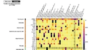
This plot is showing gene expression on the non-neuronal brain cell types (first row is BTN2A1 expression, lower rows are selected marker genes for the cell types, values for each gene are row-scaled). Microglia and circulating immune cells (just a handful each, probably caught in the vasculature) express BTN2A1 much more highly. So it seems clear that bergmann glia are probably not the source of the high cerebellar expression.

1788111143680.webp

There was one neuronal cell type that matched microglial expression levels: VGLUT1+ cerebellum granular cells. So I think that's probably why the cerebellum is enriched in BTN2A1 expression.

1788111362262.webp
 
I think it could be extremely valuable to have immunohistochemical analysis of brain and cord tissue for BTN2A1 isoforms. I have had a quick look on the net and can only find pictures using immunoperoxidase linked to a rabbit polyclonal antibody that looks very doubtful to me. Several of the images suggest non-specific carbohydrate binding (e.g. mucus). I think there may be a need to check through a panel of monoclonals to try to find one that is clean (this is a big and standard problem for IH). Peroxidase may be good, although I would favour metal enhancement for this, or alkaline phosphatase conjugates. We also need isoform specific reagents.

I suspect the lack of images reflects a general lack of interest in BTNs, because they have not come up as doing anything very important. They likely do but it has been missed. From what I have heard BTNs are quite well expressed and immunogenicity so expression patterns should be demonstrable. RNA studies are more fashionable these days but I find them much harder to interpret.

There may be more stuff on PubMed so I will look further.
 
Just a note: the more I've dug into BTN2A1 structure the more I see inconsistencies with what was written in the Coppin et al. Schizophrenia paper. For example, they claim the MOG-like domain is only in one isoform, but that feature is actually listed on all the 4 isoforms (Q7KYR7-2, Q7KYR7-4, Q7KYR7-5, Q7KYR7-6) in the Ensembl database.

They also claim in the text:
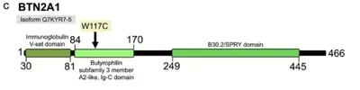
The W117C mutation occurs within the immunoglobulin V-set domain (Fig. 4c), a conserved structural motif critical for antigen recognition and immune cell-cell interactions (Barclay, 2003; Li et al., 2023).
In contrast, isoform Q7KYR7–5 (466 amino acids) exhibits a completely different domain organization. The W117C mutation in this isoform lies within the N-terminal immunoglobulin V-set domain (residues 30–84), a structurally conserved motif critical for antigen recognition and initial ligand binding in immune receptor function (Barclay, 2003; Li et al., 2023; Ramos-Miguel et al., 2015)
But from the figure from their paper that I showed earlier, it's not in the V-set domain:
1788189951690.webp
In fact both of their identified mutations are in the region that's in every isoform of the protein. Between this and the unfounded speculation about cerebellum/innate immune specific compartmentalization of isoforms (which they could have easily checked the same way I did, given the paper came out in 2026 and this GTEx resource has been available for a while), I don't think this paper is a reliable source of information for anything about BTN2A1 isoforms.
 
Last edited:
Back
Top Bottom