Loss of vesicular monoamine transporter 2 in striatum of long COVID and relationship to neuropsychiatric symptoms, 2026, Liu et al

It would be very interesting to see a study set up where LC patients (under 2 years duration?) are followed and have these scans before and after recovery.

Also I think its significant that people with parkinsons don't report PEM, and people with MECFS/LC don't have the core symptoms of Parkinsons. Obviously the hypothesis the authors propose for this finding is scary but I agree with the people here who say neuron loss doesn't fit with the clinical picture.

For example in the daratumumab pilot, we see five patients going into remission, for whatever reason (hopefully the drug). In the case of the one we know most about, she was partially bedbound at times and needed a wheelchair to leave the house. Now she is back at work and exercising and has been a few years. I don't think it likely that similar remissions would happen in five out of ten patients selected by top MECFS researchers if the cause of MECFS was permanent dopaminergic neuron loss. And we see a lot of recovery stories where people improve from moderate or severe to mild or go into remission online, whatever they happen to attribute it too.

But scary interpretation aside this finding
would fit well with some stuff we've been thinking about the last few months. I'm also interested in this link to the Hanson paper that @mariovitali has found.

It is notable and lamentable that this study does not once mention MECFS or PEM. I really hate this habit of some long covid researchers to attempt to solve long covid without engaging with or acknowledging MECFS at all. It just comes across as cowardice - not wanting their work to be associated with us. But this very silence majorly weakens the work of those who ignore MECFS.

I am glad that this team has a treatment pathway in mind. I wonder what the result of this treatment trial would be if the people in this thread are correct and they are wrong i.e. this marker is not indicating neuron loss but some kind of reversible signalling issue or other non permanent change?

It seems like this paper could be the start of something big for the field, whatever the results actually indicate in terms of neuron loss vs signalling dysfunction. Obviously I very much hope it's the latter and I'm glad to see the smart people on here think that's likely.
 
But one interesting thing is that it says this study was based on their previously seeing scans showing high TSPO levels in COVID-DNP in striatal regions:

The paper says this about previous microglial and astrocyte activation findings:
Although the integrity of dopamine terminals in striatum have not been investigated in long COVID, there are several reasons why they could be vulnerable to injury. First, individuals with long COVID show elevated markers of microglial and astroglial activation in the striatum, with some investigations reporting more widespread elevations.3,6, 7, 8

Reference 3 looks like the TSPO finding described in the protocol (S4ME thread):
References 7 and 8 are also about TSPO in long COVID:
Reference 6 tested a marker of monoamine oxidase B (MAO-B):
 
Also I think its significant that people with parkinsons don't report PEM, and people with MECFS/LC don't have the core symptoms of Parkinsons. Obviously the hypothesis the authors propose for this finding is scary but I agree with the people here who say neuron loss doesn't fit with the clinical picture.

There is a feature of Parkinson's called paradoxical kinesia, where under some circumstances patients temporarily regain normal motor control and function - eg in a situation of sudden extreme danger they may be able to move as quickly as anyone else. So the brain has some way of compensating for the effects of neuron loss, temporarily.

Maybe something not entirely unrelated happens in ME/CFS, if there is a problem with certain neurons and other parts of the brain have to be recruited to compensate for it, but on a more ongoing basis.
 
Approximately 95% of VMAT2 binding in striatum is contained within dopamine releasing neurons22 so a reduction in (+)[11C]DTBZ BPND is inferred to represent a loss of dopamine releasing neurons. This interpretation is consistently applied as VMAT2 PET imaging is a well-established method to detect pattern and progression of loss of dopamine nerve terminals in neurodegenerative illnesses and prodromal states like Parkinson's Disease, progressive supranuclear palsy, and rapid eye movement disorder.
I’m getting frustrated with the interpretations that clearly go beyond the evidence. We need to stick to the specifics of what the measurements actually are, and then try to work out exactly what caused the measurement to be what it was.

So what are we actually seeing here?

They say they used a PET tracer that’s supposed to be very selective for VMAT2:
370 MBq of (+)[11C]DTBZ (±10%) was given intravenously by bolus and scanning was done with a 3-dimensional high-resolution research tomograph PET scanner for 60 min (+)[11C]DTBZ is an excellent PET radiotracer for VMAT2 because it has high brain uptake, excellent selectivity, high affinity (Ki = 1 nM), very good specific binding relative to non-displaceable binding (BPND 1.5–2.5), very good reversibility (time activity curves peak ∼5 min), excellent reliability and is modelled in humans.18, 19, 20
Let’s assume that’s true.

The primary result was that the tracer had bound to fewer places in the regions of the brains of LC patients:
The primary outcome was that (+)[11C]DTBZ BPND was lower in all regions assayed in long COVID (LME, effect of group; long COVID mean [SD], 1.60 [0.30]; control mean [SD], 1.91 [0.21]; mean difference, −0.31; 95% CI, −0.44 to −0.17, P = 0.000038; Fig. 1A and D, Table 2).
They also found no correlation between the tracer and what they call serum markers of dopamine or neuronal injury:
Correlations of (+)[11C]DTBZ BPND with blood serum markers of dopamine or neuronal injury in the long COVID group were also negligible (Supplementary Figure S4).
I’m not sure what «dopamine injury» would be, but this is what was measured:
NfL = neurofilament light chain.
HVA = homovanillic acid.
DOPAC = 3,4-Dihydroxyphenylacetic acid.
They acknowledge that the measurements might not tell us much, though:
In addition, while low-cost peripheral plasma biomarkers may offer complementary insight, their relationship to striatal dopamine measures should be interpreted cautiously. Major sources of blood dopamine include release from chromaffin cells in the adrenal medulla, the sympathetic nervous system, and decarboxylation-active cells in the kidney and the mesentery, so contributions from the striatum represent only a small fraction.28
Likewise, the absence of a relationship between peripheral NfL and striatal (+)[11C]DTBZ BPND can be reasonably explained by the fact that NfL reflects neuronal injury to both central and peripheral neurons, so the striatum may not adequately reflect the overall source. Timing of injury relative to the assay may also be important, so this may be a measure best obtained, even if logistically difficult, during acute COVID-19.29
This leaves us with the reduced VMAT2 binding from the start of the comment:
Approximately 95% of VMAT2 binding in striatum is contained within dopamine releasing neurons22 so a reduction in (+)[11C]DTBZ BPND is inferred to represent a loss of dopamine releasing neurons.
I don’t have access to reference 22, but I would be very interested in knowing what the tracer is actually binding to in these places. They say it’s dopamine releasing neurons, but is that an interpretation and is it the whole picture? Do these neurons have different functions as well? Exactly where is VMAT2 found, and exposed to the tracer?
 
Last edited:
I don’t have access to reference 22, but I would be very interested in knowing what VMAT2 is actually binding to in these places.

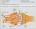
This might not be quite what you're asking but out of interest, a related graphic I found a while ago (Source):
Screenshot 2026-07-11 at 9.27.31 AM.webp
Dopamine is held (for release) in vesicles at the synapse. Wiki: "VMAT2 is an membrane protein that transports monoamines—particularly neurotransmitters such as dopamine, norepinephrine, serotonin, and histamine—from cellular cytosol into synaptic vesicles."

So I think the idea is, where there are dopamine releasing synapses, there will be VMAT2 in the dopamine vesicles' membrane. I saw VMAT2 mentioned a lot when I was reading ADHD research. But yes, I also question a bit the confidence with which some papers seems to assume lower VMAT2 definitely means one thing (when it seems like a variety of different things could cause it).
 
I don’t have access to reference 22, but I would be very interested in knowing what VMAT2 is actually binding to in these places.
I may be misunderstanding, but I think when they say VMAT2 binding, they mean the PET marker ((+)[11C]DTBZ BPND) binding to VMAT2, not measuring VMAT2 binding to other things. Although I can't access the reference either.

Here's what the protocol says:
VMAT2 is well established as a marker of dopamine neuron loss. VMAT2, is a protein in the synaptic vesicles of monoamine releasing neurons and a preferred index of dopamine releasing neurons in striatum61, 62. About 95% of the VMAT2 binding in the striatum is attributed to dopamine releasing neurons61, 62.
61 is the same as 22 in the final paper, but 62 might give some more insights.
 
All I can say given Abilify the dopamine impacting drug does help people, not cure them, maybe it is binding to a target that an AAB is so it prevents that AAB form working

I don’t think Abilify working is noise, it has helped many people until the point there is definitely some weird stuff going on with dopamine.
 
Back
Top Bottom