Preprint Persistently activated virus-specific CD8 T cells indicate antigen-driven immune aberrations in long COVID, 2026, Painter, Putrino, Iwasaki et al.

I don't know to be honest but my main worry would be about shifts in experimental conditions if batching was not well matched. Also the recovered people had had more recent infections and so the sampling may be skewed there.
Thank you. That makes sense.

I find it hard to make much of studies like this. One problem is the business about broken rules. If the data are interpreted according to normal immunological rules then you have to assume that the findings are not 'aberrations' but normal responses to peculiar circumstances.
Explain that to me a bit more. What would an example of a peculiar circumstance be?

I think I understand this bit:
If they really are aberrations then you cannot interpret them using normal rules - by definition.
So you can't interpret the aberration as indicating, say, viral persistence, is that right?
 
Last edited:
So you can't interpret the aberration as indicating, say, viral persistence, is that right??
This is something I am wondering about. They seem to be finding these T cell abnormalities in a few Polybio projects but viral persistance in the same small subgroup as in recovered controls, and COVID-19 antivirals haven't worked.

So are their findings real but pointing to something else?
 
So you can't interpret the aberration as indicating, say, viral persistence, is that right??

Yes, you cannot interpret the data that way if you think the data arise from T cells breaking the normal rules. If you have myeloma, with a massive increase in antibody to parsnip virus you cannot interpret that as indicating persistent parsnip virus because the myeloma cells are not obeying the normal rules of antibody responses - they are a malignant clone.
 
Yes, you cannot interpret the data that way if you think the data arise from T cells breaking the normal rules. If you have myeloma, with a massive increase in antibody to parsnip virus you cannot interpret that as indicating persistent parsnip virus because the myeloma cells are not obeying the normal rules of antibody responses - they are a malignant clone.
Got it. :thumbup:
 
I know that impaired cellular energy metabolism in MECFS is still debated, but I was wondering whether the following mechanism could potentially play a role.


  • Imported cellular energy metabolism could result in reduced ATP availability or a limited energy reserve in certain immune cells.
  • This might affect their ability to mount an effective antiviral response, particularly when activated or when fighting infections.
  • As a result, immune cells might struggle to control certain viruses or eliminate virus-infected cells effectively.

I came across this study on altered energy metabolism and immune profiles in MECFS:

Mapping the complexity of ME/CFS: Evidence for abnormal energy metabolism, altered immune profile, and vascular dysfunction

The study reported a reduced ATP/ADP ratio in immune cells, alongside changes in immune cell populations.

Could these findings potentially help explain the results of Iwasakis recent study, which found persistently activated viruses in a subset of people with Long COVID, showing signs of terminal effector differentiation and exhaustion?

Could altered cellular bioenergetics contribute to these T-cell abnormalities, or might persistent antigen exposure itself be sufficient to explain them?

Obviously, this is speculative, and the MECFS study does not establish a causal link between altered ATP metabolism and antiviral immune dysfunction. But I wonder whether this could be a connection worth investigating.
 
It is already a major focus across Long COVID and ME/CFS research, which is why I expect Painter and team's findings here to be replicated very soon.

Beyond the researchers who contributed to this study—like Akiko Iwasaki—US scientists such as Lisa Selin and Anayo Kohlgruber are using very similar high-resolution methods to investigate these exact hypotheses. Over here in Europe, key leaders in this space include Jacqueline Cliff and Bhupesh Prusty, all of whom have also already published excellent work on the topic and all have further studies in the pipeline. Furthermore, this research aligns directly with David Putrino's newly launched Phase II antiviral trial, providing early empirical support for the exact biological mechanisms his trial is targeting.


Good to hear. But if, with all this effort, we still don’t find the hypothesized viruses, at some point we will need to conclude they are not there. And something else is going on. Otherwise we will waste time that should be spent on working on the alternative. Which is that Iraq doesn’t have any WMD and we came all this way for nothing.
 
Good to hear. But if, with all this effort, we still don’t find the hypothesized viruses, at some point we will need to conclude they are not there. And something else is going on. Otherwise we will waste time that should be spent on working on the alternative. Which is that Iraq doesn’t have any WMD and we came all this way for nothing.
This metaphor is funny cause I’m pretty sure the top brass knew there was no WMD in Iraq to begin with
 
“In contrast, people with LC had markedly increased activation of spike-specific CD8 T cells directly ex vivo compared to controls, and this remainedstatistically significant after adjusting for age, sex, race, and SARS-CoV-2 exposure history (Fig.2A-B, Fig. S6A-B). Notably, spike-specific CD8 T cell activation was driven by a subset of peoplewith LC, and this pattern was observed across all three sites where samples were collected (Fig.S6C).”

Anyone understand how they adjust for “SARS-CoV-2 exposure”? I guess it doesn’t matter cause it was significant before adjustment?
 
Anyone understand how they adjust for “SARS-CoV-2 exposure”
Think it refers to including "Days since last vaccine/infection" and number of Sars-Cov-2 infections in the regression model. I did notice a big effect for recruitment site (being in the LIINC cohort had a bigger effect than having LC or not). That may point to technical artefacts.

1792420563729.webp
 
Okay, a couple things that stood out to me. So the plots in the main figures are for "activated CD8+ T cells", which they define as expressing at least 2 "activation markers" (the options were PD-1, CD38, CD39, Ki67, and HLA-DR).

Fig 2B:
1792442937345.webp

When they look at all CD8s, there is no difference (Fig S3A):
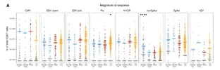
1792442534511.webp

This figure, 2D, is meant to show the distribution of markers used to call "activated" status.
1792442771717.webp
There's an additional supplemental (S6D) showing 2 more groups that were left out of the main figure:
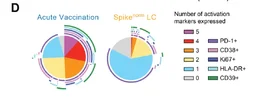
1792442833966.webp
"Spike (Act)" seems to be samples from above the dotted line in Fig 2B and "Spike (norm)" is below that line. But they kept all the recovered flattened together and splitting the LC groups this way makes it impossible to do a direct comparison on overall frequencies.

What I think is useful is to compare which activation markers are present in the acute infection condition vs. the LC samples. In all cases, the vast majority of T cells expressed PD-1 (dark purple)--that's a protein that gets upregulated once the T cell is activated and serves as a negative control mechanism (when PD-1 binds to PD-L1 it inhibits T cell responses) [edit: and usually stays upregulated through the life the T cell]

The main marker that seems to differentiate the Acute Infection condition from the LC samples is Ki67 (the dark blue)--this is a marker of proliferation and indicates that the T cell is undergoing active clonal expansion. That's virtually absent in either of the LC subsets. So really the main finding of this paper comes down to whether a T cell expressed sufficient CD38 or CD39 to be labeled as an "activated" T cell. That's where I'd really start to worry about batch effect skewing things. And there does seem to be a significant effect of recruitment site, per S6C and the plot @ME/CFS Science Blog already posted:

1792444287771.webp

They claim in the text that the difference in activated CD8+ T cells survived correction for age, sex, race and SARS-CoV-2 exposure. I guess they did not include recruitment site in the covariates. And the dotted line that they used to define "Spike (Act)/(Norm)" was the mean + 1SD for all recovered samples.

So overall I'm seeing a lot of places where technical bias and the specifics of how things were labeled may be driving this signal that was quite small to begin with. I'm willing to accept that there might be a real signal buried underneath confounders. But if it is there, I have to anticipate it's very very small.

 
Last edited:
A crucial question- did they track who did or didn't report PEM in this study?
Researchers like Iwasaki intentionally set up their Long COVID cohort criteria from the beginning around distinct subgroups and different underlying mechanisms.

They took key concepts directly from ME/CFS literature—specifically autoimmunity and smoldering herpesvirus reactivation—because they are confident in the power of modern systems immunology. Using high-dimensional tools, they knew they would have the resolution to actually map the real immunological differences between these different subgroups.

As I mentioned above, as a pre-pandemic ME/CFS patient without a clear viral trigger who experiences distinct benefits from early-phase EBV suppressants like valacyclovir and famciclovir, I believe the most crucial aspect of this paper for the broader ME/CFS community is what it demonstrates regarding EBV reactivation and chronic T-cell stimulation.

In my view, the EBV-active cohort represents the core PEM subgroup, and I am completely confident these researchers will give us more definitive answers in a relatively short timeframe. When Jackie Cliff published her findings on elevated salivary HHV-6 viral loads in ME/CFS, it took Akiko Iwasaki's team only about 18 months to replicate those results in her patients. Because these researchers have funds and methods that previous ME/CFS researchers could only dream of, the speed and resolution of this research is moving at a completely different level now.
 
Last edited:
Back
Top Bottom