Perturbation of effector and regulatory T cell subsets in Myalgic Encephalomyelitis/Chronic Fatigue Syndrome (ME/CFS) (2019) Karhan, Unutmaz et al

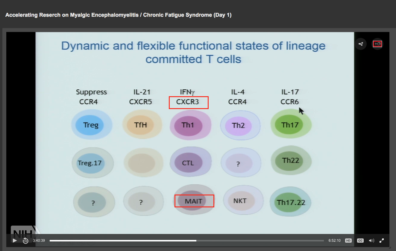
We are very excited to have just posted the full preprint of our new ME/CFS immune profiling paper on BioRxiv, which will continue to be updated following reviewer comment and peer-review. In this detailed study, we analyzed the immunological differences between ME/CFS patients and healthy controls within a large cohort and found several major differences in T cell subset frequencies and functions between the two groups.

ME/CFS is a complex and heterogeneous chronic condition that is highly debilitating and often characterized by persistent, unexplained fatigue not alleviated by rest, muscle and joint pain, sleep problems, and post-exertional malaise (PEM). There is no clear diagnostic test for the disease, so diagnosis is based largely on clinical symptoms. It’s thought that around 90% of people with ME/CFS have not been diagnosed, and up to 2.5 million Americans suffer from it, with many more suffering worldwide. This costs the US economy an estimated $17 to $24 billion annually in medical bills and lost income.
https://jaxmecfs.com/2019/12/27/jax-crc-biorxiv-preprint/
 
There was also a major difference seen in the mucosal-associated invariant T (MAIT) cells in ME/CFS patients compared to healthy controls. MAIT cells selectively respond to a broad range of bacteria, which we will discuss in more detail in a separate article. While the MAIT cell frequency did not differ between ME/CFS and controls, the functionality of these cells was profoundly different between the two groups and included a reduced production of cytokines. It is possible that these changes in Th17 and MAIT cells are associated with differences in the composition of the microbiota of the ME/CFS patients, and that a disruption in the microbiota causes chronic activation of these subsets and an exhausted state in ME/CFS patients.

Is increased intestinal permeability an important factor in the illness?

I know that during a flare up of symptoms apparently triggered by food I had a greatly increased zonulin which is I think a marker of increased intestinal permeability.

The gut problems could be a consequence of dysautonomia which may also be behind PEM and POTS.
Re: increased intestinal permeability—> we are not sure as of yet. How to prove this?
re : increased zonulin —> Who knew to test you for that? Or is it one of those test where you send your blood to a lab without going through a MD?
Lastly, I agree with your third statement, but again we do need objective measures on a sufficient sample size.
 
Re: increased intestinal permeability—> we are not sure as of yet. How to prove this?

One study found "elevated levels of some blood markers for microbial translocation in ME/CFS patients" (from the gut into the blood)
https://microbiomejournal.biomedcentral.com/articles/10.1186/s40168-016-0171-4

Another study found "increased bacterial translocation following exercise in ME/CFS patients that may account for the profound post-exertional malaise experienced by ME/CFS patients"
https://www.ncbi.nlm.nih.gov/pubmed/26683192

My zonulin test was a lucky coincidence. It was well above the norm. There may be a tenency to react more strongly with increased intestinal permeability when exposed to particular triggers, like exercise or certain foods that for some reason are problematic.

It has also been suggested that when intestinal permeability is increased, the blood brain barrier could be affected as well. There's also a story about wheat proteins leading to increased intestinal permeability even in absence of celiac disease.
 
How do I interpret Figure 9? Does this mean you could use the model that has the 10 features with highest importance and select 50% of patients correctly, and almost 0% of controls as false positive?

View attachment 9487


If yes, What is special about this 50% and could it be considered a subset? Actually I'm surprised any possible subsets are not discussed, and what about outlier patients whose results could possibly indicate an alternate disease be investigated?

Some more details : The ROC curve is typically used in Machine Learning to assess the worth of a machine learning classifier. The diagonal dashed line that appears is what we would expect when we have a random classification (so you would see all other colored lines along the dashed line). The more the line(s) lie towards the upper left corner the better. So you get the best performance with the classifier shown with the blue line. The way to read this is to see what false positive rate you expect to get for a given true positive rate (as @wigglethemouse suggests).
 
I also wanted to post this here apart from Phoenix Rising. I was in contact with Derya on April 2018. He is aware of the "Liver Injury" hypothesis but i do not know if they actually looked at the liver. I do not understand why they only look at the Gut microbiome and they are not looking at the liver (Gut + Liver associate with each other due to enterohepatic circulation).


From the NIH presentation of Derya Unutmaz at the NIH, note CXCR3 :


c5iWbw5XC2fojotwAC7Nc8gDquwmUQm6fvYVGcscEbnpIMs6mJjK_1MjWDamOwiXx5kIt1xPF9pogFI0uWfdLJUP4iXfI-6sgibiNAfUWMwqLbdtwlclXYNeAWLSayL2_lv4Tn9_


From the paper : https://www.journal-of-hepatology.eu/article/S0168-8278(16)00081-7/pdf


Title : Lights on MAIT cells, a new immune player in liver diseases


We note the connection of MAIT Cells with CXCR3 :


Upon liver inflammation, LI-MAIT cells over- express CXCR3, which is a receptor for IFN-c target genes such as CXCL9/10 and 11, as well as LFA-1 and VLA-4 integrins, suggesting the recruitment of LI-MAIT cells from the blood
 
Re: increased intestinal permeability—> we are not sure as of yet. How to prove this?
re : increased zonulin —> Who knew to test you for that? Or is it one of those test where you send your blood to a lab without going through a MD?
Lastly, I agree with your third statement, but again we do need objective measures on a sufficient sample size.

Gut permeability, there are tests for a protein which most bacteria have (bacterial skin?) - it's a cheap test. I think the test is relatively crude since it doesn't tell you which bacteria. Check out Maureen Hanson's paper(s) on bacterial translocation.
 
  • Like
Reactions: Ron
One study found "elevated levels of some blood markers for microbial translocation in ME/CFS patients" (from the gut into the blood)
https://microbiomejournal.biomedcentral.com/articles/10.1186/s40168-016-0171-4

Another study found "increased bacterial translocation following exercise in ME/CFS patients that may account for the profound post-exertional malaise experienced by ME/CFS patients"
https://www.ncbi.nlm.nih.gov/pubmed/26683192

My zonulin test was a lucky coincidence. It was well above the norm. There may be a tenency to react more strongly with increased intestinal permeability when exposed to particular triggers, like exercise or certain foods that for some reason are problematic.

It has also been suggested that when intestinal permeability is increased, the blood brain barrier could be affected as well. There's also a story about wheat proteins leading to increased intestinal permeability even in absence of celiac disease.

Gut permeability, there are tests for a protein which most bacteria have (bacterial skin?) - it's a cheap test. I think the test is relatively crude since it doesn't tell you which bacteria. Check out Maureen Hanson's paper(s) on bacterial translocation.

i have no doubt that it can be happening, but i wonder whether this is unique to our disease or whether it also occurs in other chronic diseases?
 
What association/finding did Derya Unutmaz state about T-cells expressing CXCR3 in ME patients?

I did not want to suggest that Derya found CXCR3 to be expressed, i do not know if they looked at this particular marker. I just wanted to show that there is a connection of this marker + MAIT Cells with Liver disease. Given also the following i thought it might be interesting :

=====================================================================

Title : Changes in the transcriptome of circulating immune cells of a NZ cohort with ME/CFS (2019) Sweetman et al :


From supplemental material :

r-DXr3ztBc0WAEBCz8Q255ssrbQ5krAbP6FEpGwSL5S_-cEIZ2jMTG9ewgndVd0fepLqU4C1zLdWW1_5zPdzdQyD_h4pzhITYlIBTSyceIFuSLRV3TaYtNetnIOKGXwTFIaSOY_r


https://journals.sagepub.com/doi/su...8820402/suppl_file/Supplementary_Material.pdf

=====================================================================

From NIH convention, Dr Mark Davis mentions FGF21 which is associated with Liver / Gall bladder function:


g-dw9sE1QeELs0Qc99FKOcfwiwaSsDSgsCfP-zuHftm4xkS5nvNvYAhcqTT-Q4Z8AyW4yEFnMbTckEpkayF-JXrHRLAn8qepbFSXOY_tS7wBXh9na58oE9w4LT3GmBATn7-T8sT3
 
i have no doubt that it can be happening, but i wonder whether this is unique to our disease or whether it also occurs in other chronic diseases?

My immediate thought (without thinking about this) is that gut microbiome disruptions (transition to more pathogenic species) and increased gut permeability is a feature of a number of chronic diseases - Parkinson's (?), IBS, Crohn's Disease ---. I assume that wrapped up in this is whether gut microbiome disruptions, and increased gut permeability, are cause or consequence.
I think bacterial translocation could be a central part of the disease pathology - check out Chris Armstrong's 2016 Webinar.

Also, I think increased gut permeability could be related to a switch to using amino acids for cellular energy production (related to something in the blood?) i.e. a consequence of scavenging protein from the gut to provide amino acids for cellular energy production --- SEPSIS reaction?

So I wouldn't assume that increased gut permeability is not central to the maintenance of ME. Also, I'm keen to see if leaky gut relates to something in the gut - SEPSIS response?
 
My zonulin test was a lucky coincidence. It was well above the norm. There may be a tenency to react more strongly with increased intestinal permeability when exposed to particular triggers, like exercise or certain foods that for some reason are problematic.

It has also been suggested that when intestinal permeability is increased, the blood brain barrier could be affected as well. There's also a story about wheat proteins leading to increased intestinal permeability even in absence of celiac disease.
The problem with zonulin is that it can be expressed in other tissues than the gut, so when taking a bloodtest you can't say where it originates from and where it interacts with tight junctions to loosen them up.

Regarding wheat, gliadin (the most common problematic protein in gluten) can cause increased expression of zonulin when it binds to the CXCR3 receptor on the intestinal wall. If one has to be sensitive or not for this to happen I don't know, haven't seen any studies looking at the receptors and if there are differences between those who react and those who don't.

Gut permeability, there are tests for a protein which most bacteria have (bacterial skin?) - it's a cheap test. I think the test is relatively crude since it doesn't tell you which bacteria. Check out Maureen Hanson's paper(s) on bacterial translocation.
I assume you're talking about LPS, why do you think type of bacteria is relevant once it has breached the intestinal wall?

i have no doubt that it can be happening, but i wonder whether this is unique to our disease or whether it also occurs in other chronic diseases?
It's a common enough finding in other chronic diseases, I can't really think of one where I haven't seen it mentioned.

Remember that increased permeability of the gut is a normal response to exercise also in healthy people. Is it exaggerated in us or are our bodies just unable to deal with the stress?

Several lifestyle factors can have an impact on the integrity of the intestinal wall. Vitamin D is important for barrier tissue function in general, vitamin A, zinc and glutamine the same. Other parts of the diet can affect the intestinal microbiome which again can affect the intestinal integrity (fiber for example make some bacteria produce butyrate, which increase gut integrity). Some antioxidants can also have positive effects. Sleep can affect intestinal integrity through different pathways (including the microbiome). It's all very interesting. Omega-3 fatty acids can increase expression of a protein in the gut called alkaline phosphatase, which neutralizes LPS.

I'm rambling :whistle:
 
The problem with zonulin is that it can be expressed in other tissues than the gut, so when taking a bloodtest you can't say where it originates from and where it interacts with tight junctions to loosen them up.

It was zonulin in stool.
 
I assume you're talking about LPS, why do you think type of bacteria is relevant once it has breached the intestinal wall?


Several lifestyle factors can have an impact on the integrity of the intestinal wall. Vitamin D is important for barrier tissue function in general, vitamin A, zinc and glutamine the same. Other parts of the diet can affect the intestinal microbiome which again can affect the intestinal integrity (fiber for example make some bacteria produce butyrate, which increase gut integrity). Some antioxidants can also have positive effects. Sleep can affect intestinal integrity through different pathways (including the microbiome). It's all very interesting. Omega-3 fatty acids can increase expression of a protein in the gut called alkaline phosphatase, which neutralizes LPS.

I'm rambling :whistle:

Your question "I assume you're talking about LPS, why do you think type of bacteria is relevant once it has breached the intestinal wall?" is better than my answer! I think I assumed that having more pathogenic (nasty) balance to your microbiome would mean that the translocated bacteria are likely to be bad. But yes surely increased translocation is of itself a negative.

Understanding the cause of the increased translocation is presumably fundamental.

As per @Kitty keep with the rambling!

Other distractions here - thanks for your post.
 
Bearing in mind this needs replication to be worthy of treating as fact, my preliminary comment would be the observed impairment of Th17 and MAIT cells is on the face of it at odds with the TH2 shift model where anti-bacterial responses would be expected to be elevated unless you go with the
  • "exhaustion" idea which I dont buy, or
  • homeostatic downregulation which might be a more realistic possibility or
  • pathology from an unidentified cause, in which case Th17 proliferation might be a response to pathological low activity.

I would also add that I have long been trying to explain observed episodes of cutaneous staph vulnerability in my own condition, usually regularly in mid summer when I used to get bouts of dermal oedema and cysts and zits along with pronounced pollen allergy in the context of year round recurring virus and ME CFS and while the pronounced allergy convinced me that TH2 shift idea has something going for it, the observed staph overgrowth in zits and cysts was also at odds with TH2 shift per se and might be related to this observation of decreased ability to regulate bacteria which might be TH2 shift plus something else or something like TH2 shift but not quite the same. Either way, my observations of my own condition indicate a reality where bacterial regulation is fluctuating.

I also observed a distinct vulnerability to any kind of bacterial activity in my food causing diarrhea. I attributed this to elevated sensitivity to toxins due to TH2 shift and hypersensitivity and low liver function causing the expulsion of gut contents where it was not warranted but learned to guard my food against bacteria with strict hygeine such as washing salad with detergents etc. But while the remedy may be valid the theory behind it maybe was not correct, maybe it is not sensitivity but vulnerability, due to a failure to regulate bacteria. Either way the answer is what works, avoiding bacteria in raw foods.

My conclusions are that these experimental observations might relate to the reality I experience and while its a long way from replicated and proven I cannot see any argument for disregarding these observations in relation to my own condition and once again T cell anomalies are ticking boxes. Curiouser and curiouser.

I look forward to seeing this properly verified and subgroup definitions and diagnostic tests developed, as per usual, for all the big hoorays of the last 20 years, I will believe it when I see it. Meanwhile I continue to do what works for me based on outcomes. :unsure:
 
Has anyone been able to find the supplemental table 2 referred to in the text by any chance? @Simon M ?

Machine learning analysis to identify predictive immune parameters for ME/CFS 409 410 Our immune profiling analysis identified many T cell subset parameters that were different in 411 ME/CFS patients vs healthy controls. From the total of 65 immune profile features, 40 features 412 were identified as different at a 5% false discovery rate (Supplemental Table 2). However, while 413 some of these were highly significant, given the high variability and ranges in humans for 414 immune parameters, on their own they would not have clinically relevant specificity and 415 sensitivity to discriminate patients from healthy individuals. Therefore, we decided to use a 416 classifier model using a machine learning algorithm called the random forest (RF) classifier 417 (Wang and Li, 2017).
 
Back
Top