The buspirone challenge test clearly distinguishes ME/CFS patients from healthy controls: why is it not being developed and deployed?

I don’t suppose there was any difference such as keeping the patients lying down during the whole thing in Majeed vs the others?
Majeed 1996, where no one reported nausea, doesn't seem to include the position of patients specifically for the buspirone part of the study, but for three other neuroendocrine challenges in that thesis, it says variations of this, so I assume it applies to the buspirone part too:
The subjects remained in a supine position throughout the procedure.

These are from two of the studies in which patients had greater nausea than controls. Sharpe 1996:
Subjects rested semi-supine throughout the blood sampling period and were not allowed to sleep.

The Bakheit 1992 thesis:
Subjects remained in the recumbent position during the test.

Edit: Removed mistaken interpretation. It seems like a study with nausea and a study without nausea both had participants that were fully horizontal the whole time.
 
Last edited:
Majeed 1996, where no one reported nausea, doesn't seem to include the position of patients specifically for the buspirone part of the study, but for three other neuroendocrine challenges in that thesis, it says variations of this, so I assume it applies to the buspirone part too:


These are from two of the studies in which patients had greater nausea than controls. Sharpe 1996:


The Bakheit 1992 thesis:


Edit: Removed mistaken interpretation. It seems like a study with nausea and a study without nausea both had participants that were fully horizontal the whole time.
thanks for confirming - I wasn't up atm to looking thru myself right now to find those answers. It is good to know that it was explicitly noted in each as I half expected it might not be.

I had to just check whether 'recumbent' is a broader term that 'supine'. Apparently it is and that can include the semi-supine of having a slightly reclined chair. I feel like I'm being pedantic but it feels there is the possibility that semi-supine or recumbent leaves the possibility people were in a chair without being able to have their feet up and maybe a recline of 30-60 degrees. Vs supine less than 10 degrees.

PS I'll also note I've had enough experiences in recent years where someone has 'let me' lie down but then put the back of the bed up a bit in a real bed or be on the medical couch but the back is up on that, that even if it was that and they thought it was all recumbent and nice actually it is almost a worst of both worlds because you are sort of stuck at a weird angle on something not designed to be comfy that way without decent head support at that angle (particularly when having to talk) and nothing stop you sliding, getting back ache and having to engage stomach muscles because you aren't sat up and aren't lying down. Badly reclined things that aren't really full-designed to relax with your feet up and loads of cushions behind and pillows in the right place to support head are a complete nightmare. I moved from a 'recumbent' bed into a zero gravity that supports arm and back and lifts feet etc once and the relief was visible to all in the room. I've asked to lie down in long appointments and regretted it as I'd found the couch with the back up a bit was as bad if not worse in those other ways than trying to stay in the upright chair that at least had a high back.

I don't have references but whilst certain causes of nausea you'd expect more upright to be less likely to feel sick, the OI-related and whatever the 'need to lie flat' overwhelming exhaustion wave/magnet we can all get when we hit another limit, and the length of the test I think the use of those broader, more general terms is interesting if the Majeed 1996 explicitly used supine.

I guess the other thing in my mind is method of collecting info/symptoms. I have no idea what others who have been /are in different situations than I have as far as ability to be able to report things. What I would tick on a list, particularly if it is presented like a medical questionnaire that's about eg safety for an operation and so includes what I might have had before the whatever or have all the time might be very different to what I'd see as worth reporting if someone was coming in asking for side-effects and I had to think off the top of my head.

I would also be very cautious of the context I was in and how safe I felt reporting certain things - for example I'd use terms that were 'accepted' like migraine if in a hostile situation temporarily vs talking about light sensitivity, or even just when surrounded by people who aren't going to do anything about it and feeling rubbish and so needing to speak in their language. So if I'd been stuck in a chair for hours with my feet down - I don't know details of other environmental things but was surrounded by people who would pull faces at certain more specific reports I might broaden or just leave it as nausea even tho it left it to interpretation that it was like eg those other illnesses/situations where it might be nausea, nausea rather than I'm feeling exhausted and if I was home I'd have been lying down hours ago and so I have all these symptoms that tend to relate to this as a package type nausea - and to another pwme who got it I might say 'it's OI and when I'm about to faint I feel increasingly x, y, z'.

EDIT: and of course if someone just presents you at the end with one of those endless questionnaires with boxes on then you can only tick or not tick what is on there etc. It's very hard without that extra free text when feeling awful to be able to say 'I'd be feeling this way before, but anyway if you'd stuck me in this chair for 4hrs without anything I'd probably feel more rubbish on x, y, z so who knows if it is a side-effect... where as a,b,c are definitely things I feel are 'new' or specifically related'.
 
Last edited:
But don’t let me discourage you from reaching out to a researcher if you see a connection!
I’m just exploring alternatives and throwing ideas around, nothing at all clear. So a bit at the other end of the scale from the clear focus on getting someone interested and getting a particular thing looked at.

There seems to be a large number of dopaminergic PET radiotracers for understanding what’s going in within that system so wondered if we could use them to help understand what’s is happening here.

And given it may not be the dopamine receptors themselves we’re interested in I just thinking about what else we could potentially look at when this response is happening.

There’s estrogen receptor radiotracers used in breast cancer. Could those be used here?

But also more generally just interested in what we can measure which would tell is about mechanisms rather than necessarily coming up with a mechanism and then deciding what to measure.

Some reading of folliculostellate cells has been interesting too with potential links to prolactin, ifn-gamma, il-6 and glutamine.

There’s lots of potential avenues as ever. So I’m wondering what data we could get which may lead us down one a bit more clearly.
 
Queen Square is the national centre for brain PET imaging and it looks as if they do 18F-DOPA scans. With Great ormond Street national paediatric centre across the square I suspect that nay PET techniques standardised enough to buy in will be available there. There is also likely to be a physics backup that can facilitate use of other PET labels.
 
Someone mentioned contacting Michelle James about this buspirone related PET stuff the other day. I think it's worth a shot if anyone feels knowledgeable enough about this.

If her PET paper comes out at last and there is a potential link there it might be easier to persuade her. But who knows when we'll see that.

I don't have the capacity myself right now unfortunately.
 
Queen Square is the national centre for brain PET imaging and it looks as if they do 18F-DOPA scans. With Great ormond Street national paediatric centre across the square I suspect that nay PET techniques standardised enough to buy in will be available there. There is also likely to be a physics backup that can facilitate use of other PET labels.
That’s useful to know thanks @Jonathan Edwards

I don’t understand where the costs/barriers are in these sort of studies. But it seemed worth thinking about if we already the patients, the clinicians, the support staff all there…. Trying different tracers in different people could be efficient. And casting a wide net is precisely what people didn’t do 20 years ago.

I suppose my question would be more to those neurologists or lab specialists then. Given the response we are seeing or expect to see to the drug challenge, what would it be worth measuring to help understand possible mechanisms?
 
Some reading of folliculostellate cells has been interesting too with potential links to prolactin, ifn-gamma, il-6 and glutamine.
These aee really interesting cells. And there’s loads of papers, I’m not sure where to atart but this may be of interest to peoplle. Did you come across this on your estrogen hunt @forestglip ?

(My highlighting of the abstract)

Folliculostellate Cells Determine the Susceptibility of Lactotropes to Estradiol’s Mitogenic Action, 2004, Oomizu et al

Oomizu, Souichi; Chaturvedi, Kirti; Sarkar, Dipak K.

Abstract
Estradiol is known to increase lactotropic cell proliferation, but estradiol susceptibility varies among human populations and among various strains of rats. We had reported that folliculostellate (FS) cells regulate estradiol’s mitogenic action on lactotropes; therefore, we studied their role in determining the susceptibility to estradiol in a high estradiol-responsive rat strain, Fischer 344 (F344), and in a low-responsive strain, Sprague Dawley (SD). Determination of total S-100-positive FS cells in the pituitary revealed that F344 rats have significantly more FS cells than do SD rats.

Estradiol treatment did not change the number of FS cells in both F344 and SD rats. When cotransplanted with F344 pituitaries under the kidney capsule or cocultured with F344-derived lactotropes in vitro, FS cells derived from F344 rats increased estradiol’s mitogenic action. They also increased estradiol’s mitogenic action on SD-derived lactotropes in primary cultures.

However, SD-derived FS cells failed to increase estrogen’s action on F344- or SD-derived lactotropes. The levels of basic fibroblast growth factor production and secretion by TGF-β3 and estradiol were much higher in F344-derived FS cells than in SD-derived FS cells. However, the lactotropes’ growth response to basic fibroblast growth factor was similar in both strains.

These data suggest that cell-cell interaction between FS cells and lactotropes regulates estradiol’s mitogenic action on lactotropes and also determines lactotrope susceptibility to the steroid.

Web | DOI | PMC | PDF | Endocrinology
 
There’s also a big old review here which touches o some interesting ideas of how these cells manage the pitutary axes and may deal with memory or anticipation of demand.

(This may be one for another thread of ‘things hotblack has found interesting’ rather than clogging up discussion here, I can move in the morning if needed)
Edit: thread for all things Folliculostellate cell

Renewing an old interest: Pituitary folliculostellate cells, 2021, Le Tissier et al

Le Tissier, Paul R.; Mollard, Patrice

Abstract
Anterior pituitary folliculostellate (FS) cells, first described almost 50 years ago, have a wide range of functions with respect to supporting and coordinating endocrine cell function, in particular through paracrine and gap junction‐mediated signalling.

Our previous studies identified the morphological organisation of FS cells, which mediates coordinated calcium activity throughout the homotypic FS network and allows signalling across the whole pituitary gland. It is also clear that FS cells can modify endocrine output and feedback on pituitary axes over a range of timescales.

Recently, several studies have defined FS cells as a source of anterior pituitary endocrine cell renewal, which has resulted in a renaming of FS cells as “Sox2+ve stem cells”.

Here, we highlight the broader potential of the FS cell population in fine‐tuning and coordinating pituitary axes function. In addition, we identify a need for: the definition of the possible subtypes of FS cell and their relationship with the stem cell population; the potential role of FS cells in pulsatile hormone secretion and coordination of heterotypic cell networks; and the roles that FS cells may play in both early‐life programming of pituitary axes and in memory, or anticipation, of demand.

Further studies of FS cells may demonstrate the fundamental importance of this cell type and its potential as a therapeutic target to correct pituitary gland dysfunction, one of which is stem cell therapy. Clearly, a thorough understanding of all of these interactions and relationships of FS and endocrine cells is required whatever therapeutic use is suggested by their various roles.

Web | DOI | PMC | PDF | Journal of Neuroendocrinology
 
Last edited:
I took a tour of the Google Scholar results for "prolactin fenfluramine" and found a few somewhat interesting things. I mostly haven't read the full text of these, and am going by the abstracts.


The increase in prolactin following fenfluramine was found to be completely blocked by pre-treatment with ritanserin (selective antagonist of 5-HT2A and 5-HT2C) or amesergide (selective antagonist of 5-HT2A, 5-HT2B, and 5-HT2C).

Pre-treatment with pindolol (antagonist for 5-HT1A and β-adrenergic receptors) found attenuation or no effect on fenfluramine-induced prolactin response in different studies.

Pre-treatment with amperozide (5-HT1A 5-HT2 antagonist) did not alter fenfluramine's effect on prolactin.

[Edit: I mixed up the receptor for amperozide, so the interpretation is less clear than I wrote here.]

I'm not an expert on all these receptors, but it seems like this might be decent evidence that the 5-HT1A is not involved in fenfluramine's effect on prolactin, or at least only has a minor role, and that instead one of the 5-HT2 receptors might be a receptor involved in its effect.

The fenfluramine and buspirone studies led Cleare et al. to measure levels of the 5-HT1A receptor in the brains of those with chronic fatigue syndrome. They found somewhat lower levels throughout the brain, which was the opposite direction of what was expected, and it seems that line of research went no further after that. I wonder if the data here would suggest that other 5-HT receptors might be better candidates for investigating the possibility of serotonin receptor abnormalities.

Also, don't take my word for the selectivity of the drugs. I quickly copied that from Wikipedia, but it seems to align with how the abstracts of these papers describe the drugs.

Pharmacological characterization of serotonin receptors involved in the control of prolactin secretion (1989) (Rats)
Ritanserin (200 μg/kg i.p.), a specific antagonist of 5-HT2 receptors, administered 1 h before the administration of d-fenfluramine or quipazine, completely prevented the PRL-releasing effect of these drugs.

Ritanserin attenuates anorectic, endocrine and thermic responses tod-fenfluramine in human volunteers (1993)
ritanserin abolished the [d-fenfluramine] induced rise in PRL

5-HT2a/2c receptor blockade by amesergide fully attenuates prolactin response tod-fenfluramine challenge in physically healthy human subjects (1996)
pre-treatment with amesergide completely blocked the prolactin (PRL) response tod-FEN challenge in all subjects.

Involvement of the 5-HT2 receptor in the 5-HT receptor-mediated stimulation of prolactin release (1994) (Rats)
amperozide did not attenuate d-fenfluramine-elicited prolactin release

Evidence for 5-hydroxytryptamine1A receptor involvement in the control of prolactin secretion in man (1995)
Pindolol pretreatment attenuated the (+)-fenfluramine-induced increase in prolactin concentrations

Effect of pindolol on the prolactin response to d-denfluramine (1995)
Pindolol pretreatment lowered baseline PRL levels but, when this effect was taken into account, did not significantly attenuate the PRL response to d-FEN.

Edit: I accidentally wrote 5-HT1A antagonist instead of 5-HT2 antagonist for amperozide.
 
Last edited:
4 weeks of treatment with the dopamine antagonist haloperidol led to higher baseline prolactin, but no change in prolactin response to fenfluramine (Mohr et al., 1999). I'm not sure whether or not it can be concluded from this that dopamine is not involved in the pathway that fenfluramine uses to increase prolactin.

Studies of other serotonin drugs suggest that dopamine receptors are a step in the pathway these drugs use to increase prolactin, quoted below, but maybe fenfluramine binds to different receptors and thus takes a different pathway.

Partial Characterization of a Neurotransmitter Pathway Regulating the in vivo Release of Prolactin (1992)
Bromocriptine [dopamine antagonist] blocked the prolactin secretion induced [...] by each of the above agonists [including the serotonin 1A (5-HT1A) agonist 8-hydroxy-2-(di-n-propylamino)tetralin (8-OH-DPAT)].

Effects of the serotonin agonist, quipazine, on luteinizing hormone and prolactin release: Evidence for serotonin-catecholamine interactions (1984)
Quipazine [serotonin agonist] (15 mg/kg, ip) significantly reduced LH and increased PRL when administered to ovariectomized rats. Associated with these changes, the depletion of dopamine seen after synthesis inhibition with α-methyl tyrosine was reduced by quipazine in the caudate nucleus and median eminence, suggesting a depression of dopaminergic activity.


The following study found a correlation between platelet MAO-B activity and prolactin response to DL-fenfluramine. I don't know what it could mean, but I thought it was worth noting.

Platelet monoamine oxidase B (MAO-B) activity and its relationship to DL-fenfluramine-induced prolactin response in healthy men (2006)
 
Pre-treatment with amperozide (5-HT1A 5-HT2 antagonist) did not alter fenfluramine's effect on prolactin.
Noting that I didn't read it carefully, and wrote the wrong receptor here.

Paper about amperozide:
Amperozide possessed a high affinity to the 5-HT2 receptors (Ki= 16.5±2.1 nM) and a moderate affinity to α1-adrenergic receptors of rat cerebral cortical membranes (Ki=172±14 nM). [...] The affinity of amperozide for striatal and limbic dopamine D2 receptors was low [...] The affinity for several other rat brain receptors such as 5-HT1A, α2-adrenergic, dopamine D1 muscarinic M1 and M2, opiate sigma and β2-adrenergic was low.

The paper about it's effect on fenfluramine-prolactin release said:
These results, along with the finding that the selective 5-HT2 receptor antagonist, amperozide, did not block a fenfluramine-induced increase in plasma prolactin, indicate that the 5-HT1c receptor may be of greater importance than the 5-HT2 receptor in the physiological regulation of prolactin secretion.
 
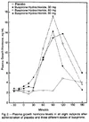
Two other things we could check are if the 30 minute intervals which are also on the growth hormone plot make sense (is the peak consistent with other studies?)
I found one study of growth hormone response that includes a plot:

The Effect of Buspirone on Prolactin and Growth Hormone Secretion in Man (Meltzer et al., 1983)

Peak for growth hormone after buspirone at around 90-120 minutes:
1791062221061.webp

In the Sharpe 1996 ME/CFS study, the peak was at around 120-150 minutes.
1791062291029.webp

So this seems fairly consistent, and adds some evidence that the times for the prolactin plot are correct.
 
but if I had to choose only one specific study to start with, it'd be a trial both re-testing buspirone (to verify that these individuals have the same thing seen in earlier studies) and also testing domperidone in the same individuals (to see if the finding specifically relates to dopamine levels/D2 receptors, something else within lactotrophs, or if it's something more upstream).
I do like the sound of that. If it was confirmed there’s something going on around lactotrophs maybe we can get endocrinologists interested. And they can look at FS cells :D
No, I haven't. Maybe there's something there.
There may be nothing. But there may be something!

FS cells could potentially tie in with changes in numbers of or perhaps more likely sensitivity and responsiveness of lactotrophs. And in response to immune signals, maybe even changes over time and at particular stages of life. And it would be another funny way in which people have been close to being in the right path for years but not looking at things the right way. There’s a story which could be weaved around them from the reading I’ve done I think. Not that that means much but hey, if it gets more researchers interested and prodding around…
 
Regarding estrogens as an explanation for increased prolactin response, the problem with that hypothesis is that it's somewhat consistent that the studies that show an increased prolactin response for a sex hormone-related reason also show increased baseline prolactin. In contrast, ME/CFS does not seem to present with changes in baseline prolactin.

For example, going through the studies I cited about this potential sex hormone involvement in my review:
(I've made the reference numbers in the quotes link directly to the papers.)

Females have larger prolactin response than males
The clearest connection of prolactin response findings to the sex bias in ME/CFS comes from observations that females tend to have larger prolactin responses to various neuroendocrine probes than males. For example, healthy females have a larger prolactin response to buspirone [28, 35]. Similar sex differences in prolactin response magnitude have been observed after administration of d-fenfluramine [106], thyrotropin-releasing hormone (TRH) [107], phenothiazine [108], and possibly chlorpromazine [109].

Apart from references 35 (baseline difference not reported) and 108 (non-significant difference), all the references above found higher baseline prolactin in females.

Prolactin response varies through menstrual cycle
O’Keane et al. observed that while baseline prolactin levels in healthy females stayed relatively steady throughout the menstrual cycle, prolactin response to d-fenfluramine was highest at mid-cycle, followed by the luteal, then follicular phase, which mirrored the differing amounts of circulating estradiol measured at different phases [110]. Prolactin response to buspirone in females was found to be larger during the luteal phase than during the follicular phase or at mid-cycle [35]. Prolactin response to TRH may be somewhat larger during the follicular [periovulatory] phase [111], though findings are inconsistent [112].
(Note that I wrote follicular phase for TRH response, but @EndME notified me that was incorrect and should say "periovulatory". I'll fix in a future version. [Edit: The reason was I mixed up periovulatory and preovulatory.])

For 110 and 111, baseline prolactin did not differ between menstrual phases. 35 didn't report baseline prolactin levels in females. For 112, quoted below, there might have been a couple dips in prolactin, after menses and after ovulation.
No difference was observed between basal PRL concentrations during the proliferative and secretory phases of the menstrual cycle although there was a consistent dip after menses and immediately after ovulation.
[On smaller subset of women tested every day] While the standard errors were too wide to make the values statistically significant, again prolactin appears to decline soon after menses and for a second time just beyond mid-cycle.

Other studies: For Bäckström 1982, it looks like prolactin was lowest at early-mid luteal phase.

Franchimont 1976: Lowest during follicular phase.
However, for each of the women it was confirmed that the mean levels of prolactin during the ovulatory phase were always greater than those during the follicular phase and the mean level during the luteal phase was greater than that of the follicular phase in twelve of fourteen cases and lower than that in the ovulatory phase in ten of fourteen. For the whole group these differences were highly statistically significant.

Tanner 2011: Lowest during follicular phase.
Within premenopausal women, the prolactin median and upper limits were significantly higher in ovulatory phase compared to follicular/non-cycling and luteal phases and in luteal phase compared to follicular/non-cycling phase.

Reference 111 says findings from studies have been inconsistent:
a cyclical pattern of serum prolactin, similar to that of endogenous oestrogens, has been reported during the normal menstrual cycle (Robyn et al., 1973), but this is questioned by other authors (Jaffe et al., 1973; McNejlly & Chard, 1974; Fournier et al., 1974).

So it's not consistent, but some studies found variation in baseline prolactin throughout the menstrual cycle. Though it's interesting that in the study of prolactin response to d-fenfluramine at different phases, baseline prolactin did not differ while prolactin response to the drug did.

Direct effect of estradiol on prolactin response
TRH-induced prolactin response was seen to be substantially larger in females taking a short course of exogenous estradiol, as well as after subsequent short-term combined contraceptive use, but not in those taking long-term combined contraceptives [111]. In male rats, a prolactin response to TRH was only detectable when the rats had previously been administered estradiol [113]. Exogenous estradiol causes an increased prolactin response to ghrelin in postmenopausal women [114]. Administration of estradiol to ovariectomized rats resulted in substantially larger prolactin responses to the dopamine antagonist, thioproperazine [115].

For 111, in females taking sequential contraceptives (16 days estradiol, then 7 days estradiol+progestogen), prolactin response to TRH was higher than in normally cycling females at the end of both phases of treatment, but baseline prolactin was only higher during the estradiol-only phase.

However, long term combined estradiol+progestogen that did not follow an estradiol-only phase did not appear to cause increased prolactin response.

For 113, where prolactin response to TRH was only detectable in male rats after estradiol treatment, the paper doesn't give the baseline prolactin for the rats that weren't treated. There were two other experiments in the study, and it seems like in one, estradiol increased baseline prolactin, while in the other, it did not. Maybe it's related to dose or specific form of estradiol (one study used 25 mcg 17B-estradiol, the other used 50 mcg estradiol-benzoate).

For 114, where estradiol valerate increased prolactin response to ghrelin in post-menopausal females, baseline prolactin was also increased.

For 115, where estradiol increased prolactin response to thioproperazine, they don't describe baseline prolactin and it's hard to figure out the values from the plot.

So the closest analog to the ME/CFS findings seems to be at the end of a cycle of women taking 16 days estradiol-only, then 7 days estradiol+progestogen, where baseline prolactin was similar to in normally cycling women, but prolactin response was significantly larger. (Though this is prolactin response to TRH, which has not been tested in ME/CFS.) (Reymond & Lemarchand-Béraud, 1976).

But long-term estradiol+progestogen without an estradiol-only phase did not cause increased prolactin response. Here's their discussion about the difference between the two treatments:
Sequential oral contraceptives contain higher concentrations of oestrogens than the combined pills (100 ug ethinyl oestradiol v. 50 ug). Moreover, the oestrogen phase of the treatment with sequential contraceptives, ensuring continuous exposure of the pituitary for 16 days, may result in a modification of the ‘set point’ of prolactin release, which persists even when progestogens are added subsequently to oestrogens.
So maybe estradiol increases both baseline prolactin and prolactin response, then subsequent progestegen brings baseline prolactin back to normal, while prolactin response stays elevated for some time longer.

Though it's not clear why something like what is seen after short-term but not long-term combined contraceptives would be permanently present in ME/CFS. And it's only based on one study of the contraceptive treatment, so the confidence about the contraceptive finding itself is not based on a lot of evidence.
 
Last edited:
So maybe estradiol increases both baseline prolactin and prolactin response, then subsequent progestegen brings baseline prolactin back to normal, while prolactin response stays elevated for some time longer.

Maybe this would track with the idea that one of the ways estradiol raises prolactin is just by causing lactotrophs to proliferate (a change I imagine would take longer to dial back):
Grattan said:
In the pituitary gland, estradiol is a major stimulator of prolactin secretion, although this is principally through a classical genomic regulation of prolactin gene expression, by increasing the number of lactotrophs (Takahashi et al. 1984, Scully et al. 1997, Kansra et al. 2005, 2010, Nolan & Levy 2009) and by modifying lactotroph responsiveness to other regulators [...]
 
Maybe this would track with the idea that one of the ways estradiol raises prolactin is just by causing lactotrophs to proliferate (a change I imagine would take longer to dial back):
So still large number of lactotrophs, but the genes within each lactotroph have been reprogrammed to release a normal level of prolactin. But when they get a stimulus, the increase is still larger due to the many lactotrophs? I guess something like that could be happening in ME/CFS.

Alternatively, the lack of change in baseline prolactin might be a sign it's not related to sensitized lactotrophs or estrogens, and is something further upstream in the CNS. I think it'd probably be a bigger challenge to figure out what to do next if that was the case. Maybe continuing to test lots of probes, or PET scans on all sorts of receptors.

It seems like the bulk of prolactin response research in other conditions concluded that things were abnormal in the CNS, like serotonin receptor hypersensitivity. But after hundreds of studies in various conditions, it doesn't seem like findings in any conditions led to any meaningful insights. Happy for someone to correct me on that.

However, it also doesn't seem like the findings were very consistent in many other conditions. Maybe in the testicular failure conditions, increased prolactin response was fairly consistent (though mostly from one lab). That one and ovarian failure were the only conditions I can remember where they speculated that the reason might be related to estrogens. But nothing came of that either, as far as I know.

If increased prolactin response to buspirone continues to be consistently replicated in ME/CFS, it might be one of the few conditions where abnormal prolactin response is consistent and predictable enough where it's possible to gain insights from it.
 
I think it could work with an increased number of lactotrophs or greater responsiveness of those lactotrophs for some reason.

With largely steady state levels the balance between supressjon from dopamine and everything else would be just fine. It would only be with sharp changes in levels that the change is amplified and therefore you get the higher peak. The systems to bring things back into balance as well as keep them level are fine, they just may not compensate immediately to sharp changes in prolactin secretion.

I’m thinking of behaviour (particularly gain/amplification) in electrical circuits here as that’s something I’m a bit more familiar with, but I think the behaviour matches.

I wonder if the dosage helps show this? We need to ensure an abrupt change rather than a slow drift? Maybe more reason to look at dose response? Something to think about with domperidone too?
 
Last edited:
Back
Top Bottom