Not sure if this is the right place to put this, but I wanted to consolidate a couple thoughts that span multiple threads. I was looking into the genes driving eMSN enrichment in Paolo's analysis. The leading edge genes from the top hit were, in order: DNMT3A / PPP2R2A / SYNGAP1 / DLGAP4 / CACNA2D3 / ZC3H13 / CAMK2A / PTPN11 / PDYN / CDR2 / BAIAP2
I should note that all of these hits were a very weak signal in DecodeME. But with the exception of BAIAP2, all were in the core list of Zhang et al. hits (DNMT3A being the #1 hit and several others being top 20)
Considering that Zhang et al. is a rare gene analysis which focused on predicted loss of function genes, I did some more digging on what effects loss of function in those genes might have (focusing on effects that aren't necessarily developmental and can manifest later in life).
I found this paper for DMNT3A:
Deleterious somatic mutations in DNA methyltransferase 3 alpha (DNMT3A) and TET mehtylcytosine dioxygenase 2 (TET2) are associated with clonal expansion of hematopoietic cells and higher risk of cardiovascular disease (CVD). Here, we investigated roles of DNMT3A and TET2 in normal human monocyte-derived macrophages (MDM), in MDM isolated from individuals with DNMT3A or TET2 mutations, and in macrophages isolated from human atherosclerotic plaques. We found that loss of function of DNMT3A or TET2 resulted in a type I interferon response due to impaired mitochondrial DNA integrity and activation of cGAS signaling. DNMT3A and TET2 normally maintained mitochondrial DNA integrity by regulating the expression of transcription factor A mitochondria (TFAM) dependent on their interactions with RBPJ and ZNF143 at regulatory regions of the TFAM gene. These findings suggest that targeting the cGAS-type I IFN pathway may have therapeutic value in reducing risk of CVD in patients with DNMT3A or TET2 mutations.
Obviously mtDNA/interferon is the favorite pet theory of your local graduate student, so I assumed my attention was just being caught by what I was already looking for.
But interestingly, a second gene from this list (PPP2R2A) was associated with the
exact same thing:
PP2A B55α, a regulatory subunit of protein phosphatase 2 (PP2A), is underexpressed in greater than 40% of non–small cell lung cancer (NSCLC) cases due to loss of heterozygosity of PPP2R2A, the gene encoding this protein. Given that low PPP2R2A ...

pmc.ncbi.nlm.nih.gov
PP2A B55α, a regulatory subunit of protein phosphatase 2 (PP2A), is underexpressed in greater than 40% of non–small cell lung cancer (NSCLC) cases due to loss of heterozygosity of PPP2R2A, the gene encoding this protein. Given that low PPP2R2A expression correlates with poor prognosis, treating PPP2R2A-deficient NSCLC represents an unmet medical need. Here, we show that PPP2R2A knockdown or its heterozygosity (PPP2R2A+/–) increases cytosolic DNA, leading to cGAS-STING-type I IFN pathway activation. PPP2R2A deficiency results in elevated expression of immune checkpoint protein PD-L1 via GSK-3β- and STING-dependent mechanisms. PPP2R2A+/– cancer cells have enhanced sensitivity to PD-L1 blockade in a mouse model of lung cancer due to modulation of the tumor immune microenvironment, resulting in increased NK cells and reduced infiltration and function of Tregs. Consequently, PD-L1 antibody treatment increases CD8+ T infiltration and activity, especially in tumors with PPP2R2A heterozygosity. Furthermore, systemic or Treg-specific IFNAR1 blockade reduces the efficacy of PD-L1 blockade in PPP2R2A+/– tumors. Patients with NSCLC with a low PPP2R2A/PD-L1 ratio respond better to immune checkpoint blockade (ICB). These findings underscore the therapeutic potential of ICB in treating PPP2R2A-deficient NSCLC and suggest that PPP2R2A deficiency could serve as a biomarker for guiding ICB-based therapies.
CAM2KA is part of the CAMKII complex, which mediates an important calcium-responsive signaling cascade. I couldn't link loss of function of CAM2KA to mtDNA release, though post-translational modifications on another protein that CAM2KA interacts with (in the CAMKII complex) have been associated with it in a heart disease context:
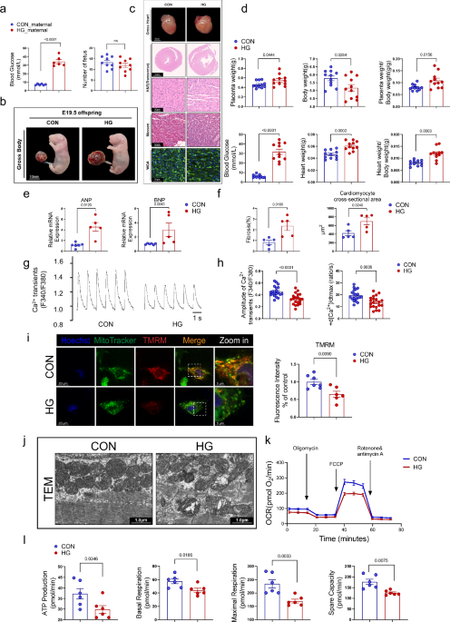
The study demonstrates that maternal hyperglycemia activates CaMKIIδ via O-GlcNAcylation, leading to mitochondrial DNA release. Targeting this pathway alleviates cardiac remodeling and identifies circulating mtDNA as a potential biomarker of fetal cardiac injury.

www.nature.com
So several genes that are upregulated in eMSNs and picked up in genetic analyses have direct links to the same biological process.
These examples are in heart tissue and immune cells, so the connection to eMSNs wasn't entirely clear. But given that MSNs appear to have
different calcium handling than other neurons and mtDNA/RNA release is a calcium-responsive phenomenon, I wanted to see if there was any link to this phenomenon happening in neurons. Turns out MSNs, the cell type most affected in Huntington's, showed distinct mtRNA and DNA release in both mouse models and human samples:
The mechanisms by which mutant huntingtin (mHTT) leads to neuronal cell death in Huntington’s disease (HD) are not fully understood. To gain new molecular insights, we used single nuclear RNA sequencing (snRNA-seq) and translating ribosome affinity purification (TRAP) to conduct transcriptomic analyses of caudate/putamen (striatal) cell type-specific gene expression changes in human HD and mouse models of HD. In striatal spiny projection neurons, the most vulnerable cell type in HD, we observe a release of mitochondrial RNA (mtRNA) (a potent mitochondrial-derived innate immunogen) and a concomitant upregulation of innate immune signaling in spiny projection neurons. Further, we observe that the released mtRNAs can directly bind to the innate immune sensor protein kinase R (PKR). We highlight the importance of studying cell type-specific gene expression dysregulation in HD pathogenesis and reveal that the activation of innate immune signaling in the most vulnerable HD neurons provides a novel framework to understand the basis of mHTT toxicity and raises new therapeutic opportunities.
We've talked about huntington's a few times on the forum, and one thing that always struck me as interesting about it was the late in life onset. It makes me wonder whether there's something that happens in this neuron population starting in midlife. Probably an extra reason to make sure age of onset is catalogued in any future rare variant analyses in ME/CFS.
Also, this study did find a population of FOXP2+ neurons that are probably eMSNs, and surprisingly they were the only cell population that actually showed less mtRNA release during active huntington's disease compared to controls (the 2 other MSN populations had the strongest upregulation). It was a small sample size and I'm not sure from the supplemental material if the same finding was corroborated in their mouse model. But if it is real, it makes me curious whether there is something specific about eMSNs that normally protects them from mtRNA/DNA release, and whether the genetic hits in DecodeME might point to that mechanism being bypassed.
I should also note that the Huntington's paper didn't establish mtRNA/DNA release was directly the cause of cell death in that disease. It might just be a contributing factor or something that happens downstream when HTT accumulation is already stressing the cell.