Loss of vesicular monoamine transporter 2 in striatum of long COVID and relationship to neuropsychiatric symptoms, 2026, Liu et al

It would be very interesting to see a study set up where LC patients (under 2 years duration?) are followed and have these scans before and after recovery.

Also I think its significant that people with parkinsons don't report PEM, and people with MECFS/LC don't have the core symptoms of Parkinsons. Obviously the hypothesis the authors propose for this finding is scary but I agree with the people here who say neuron loss doesn't fit with the clinical picture.

For example in the daratumumab pilot, we see five patients going into remission, for whatever reason (hopefully the drug). In the case of the one we know most about, she was partially bedbound at times and needed a wheelchair to leave the house. Now she is back at work and exercising and has been a few years. I don't think it likely that similar remissions would happen in five out of ten patients selected by top MECFS researchers if the cause of MECFS was permanent dopaminergic neuron loss. And we see a lot of recovery stories where people improve from moderate or severe to mild or go into remission online, whatever they happen to attribute it too.

But scary interpretation aside this finding
would fit well with some stuff we've been thinking about the last few months. I'm also interested in this link to the Hanson paper that @mariovitali has found.

It is notable and lamentable that this study does not once mention MECFS or PEM. I really hate this habit of some long covid researchers to attempt to solve long covid without engaging with or acknowledging MECFS at all. It just comes across as cowardice - not wanting their work to be associated with us. But this very silence majorly weakens the work of those who ignore MECFS.

I am glad that this team has a treatment pathway in mind. I wonder what the result of this treatment trial would be if the people in this thread are correct and they are wrong i.e. this marker is not indicating neuron loss but some kind of reversible signalling issue or other non permanent change?

It seems like this paper could be the start of something big for the field, whatever the results actually indicate in terms of neuron loss vs signalling dysfunction. Obviously I very much hope it's the latter and I'm glad to see the smart people on here think that's likely.
 
But one interesting thing is that it says this study was based on their previously seeing scans showing high TSPO levels in COVID-DNP in striatal regions:

The paper says this about previous microglial and astrocyte activation findings:
Although the integrity of dopamine terminals in striatum have not been investigated in long COVID, there are several reasons why they could be vulnerable to injury. First, individuals with long COVID show elevated markers of microglial and astroglial activation in the striatum, with some investigations reporting more widespread elevations.3,6, 7, 8

Reference 3 looks like the TSPO finding described in the protocol (S4ME thread):
References 7 and 8 are also about TSPO in long COVID:
Reference 6 tested a marker of monoamine oxidase B (MAO-B):
 
Also I think its significant that people with parkinsons don't report PEM, and people with MECFS/LC don't have the core symptoms of Parkinsons. Obviously the hypothesis the authors propose for this finding is scary but I agree with the people here who say neuron loss doesn't fit with the clinical picture.

There is a feature of Parkinson's called paradoxical kinesia, where under some circumstances patients temporarily regain normal motor control and function - eg in a situation of sudden extreme danger they may be able to move as quickly as anyone else. So the brain has some way of compensating for the effects of neuron loss, temporarily.

Maybe something not entirely unrelated happens in ME/CFS, if there is a problem with certain neurons and other parts of the brain have to be recruited to compensate for it, but on a more ongoing basis.
 
Approximately 95% of VMAT2 binding in striatum is contained within dopamine releasing neurons22 so a reduction in (+)[11C]DTBZ BPND is inferred to represent a loss of dopamine releasing neurons. This interpretation is consistently applied as VMAT2 PET imaging is a well-established method to detect pattern and progression of loss of dopamine nerve terminals in neurodegenerative illnesses and prodromal states like Parkinson's Disease, progressive supranuclear palsy, and rapid eye movement disorder.
I’m getting frustrated with the interpretations that clearly go beyond the evidence. We need to stick to the specifics of what the measurements actually are, and then try to work out exactly what caused the measurement to be what it was.

So what are we actually seeing here?

They say they used a PET tracer that’s supposed to be very selective for VMAT2:
370 MBq of (+)[11C]DTBZ (±10%) was given intravenously by bolus and scanning was done with a 3-dimensional high-resolution research tomograph PET scanner for 60 min (+)[11C]DTBZ is an excellent PET radiotracer for VMAT2 because it has high brain uptake, excellent selectivity, high affinity (Ki = 1 nM), very good specific binding relative to non-displaceable binding (BPND 1.5–2.5), very good reversibility (time activity curves peak ∼5 min), excellent reliability and is modelled in humans.18, 19, 20
Let’s assume that’s true.

The primary result was that the tracer had bound to fewer places in the regions of the brains of LC patients:
The primary outcome was that (+)[11C]DTBZ BPND was lower in all regions assayed in long COVID (LME, effect of group; long COVID mean [SD], 1.60 [0.30]; control mean [SD], 1.91 [0.21]; mean difference, −0.31; 95% CI, −0.44 to −0.17, P = 0.000038; Fig. 1A and D, Table 2).
They also found no correlation between the tracer and what they call serum markers of dopamine or neuronal injury:
Correlations of (+)[11C]DTBZ BPND with blood serum markers of dopamine or neuronal injury in the long COVID group were also negligible (Supplementary Figure S4).
I’m not sure what «dopamine injury» would be, but this is what was measured:
NfL = neurofilament light chain.
HVA = homovanillic acid.
DOPAC = 3,4-Dihydroxyphenylacetic acid.
They acknowledge that the measurements might not tell us much, though:
In addition, while low-cost peripheral plasma biomarkers may offer complementary insight, their relationship to striatal dopamine measures should be interpreted cautiously. Major sources of blood dopamine include release from chromaffin cells in the adrenal medulla, the sympathetic nervous system, and decarboxylation-active cells in the kidney and the mesentery, so contributions from the striatum represent only a small fraction.28
Likewise, the absence of a relationship between peripheral NfL and striatal (+)[11C]DTBZ BPND can be reasonably explained by the fact that NfL reflects neuronal injury to both central and peripheral neurons, so the striatum may not adequately reflect the overall source. Timing of injury relative to the assay may also be important, so this may be a measure best obtained, even if logistically difficult, during acute COVID-19.29
This leaves us with the reduced VMAT2 binding from the start of the comment:
Approximately 95% of VMAT2 binding in striatum is contained within dopamine releasing neurons22 so a reduction in (+)[11C]DTBZ BPND is inferred to represent a loss of dopamine releasing neurons.
I don’t have access to reference 22, but I would be very interested in knowing what the tracer is actually binding to in these places. They say it’s dopamine releasing neurons, but is that an interpretation and is it the whole picture? Do these neurons have different functions as well? Exactly where is VMAT2 found, and exposed to the tracer?
 
Last edited:
I don’t have access to reference 22, but I would be very interested in knowing what VMAT2 is actually binding to in these places.

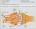
This might not be quite what you're asking but out of interest, a related graphic I found a while ago (Source):
Screenshot 2026-07-11 at 9.27.31 AM.webp
Dopamine is held (for release) in vesicles at the synapse. Wiki: "VMAT2 is an membrane protein that transports monoamines—particularly neurotransmitters such as dopamine, norepinephrine, serotonin, and histamine—from cellular cytosol into synaptic vesicles."

So I think the idea is, where there are dopamine releasing synapses, there will be VMAT2 in the dopamine vesicles' membrane. I saw VMAT2 mentioned a lot when I was reading ADHD research. But yes, I also question a bit the confidence with which some papers seems to assume lower VMAT2 definitely means one thing (when it seems like a variety of different things could cause it).
 
I don’t have access to reference 22, but I would be very interested in knowing what VMAT2 is actually binding to in these places.
I may be misunderstanding, but I think when they say VMAT2 binding, they mean the PET marker ((+)[11C]DTBZ BPND) binding to VMAT2, not measuring VMAT2 binding to other things. Although I can't access the reference either.

Here's what the protocol says:
VMAT2 is well established as a marker of dopamine neuron loss. VMAT2, is a protein in the synaptic vesicles of monoamine releasing neurons and a preferred index of dopamine releasing neurons in striatum61, 62. About 95% of the VMAT2 binding in the striatum is attributed to dopamine releasing neurons61, 62.
61 is the same as 22 in the final paper, but 62 might give some more insights.
 
All I can say given Abilify the dopamine impacting drug does help people, not cure them, maybe it is binding to a target that an AAB is so it prevents that AAB form working

I don’t think Abilify working is noise, it has helped many people until the point there is definitely some weird stuff going on with dopamine.
 
All I can say given Abilify the dopamine impacting drug does help people, not cure them, maybe it is binding to a target that an AAB is so it prevents that AAB form working

I don’t think Abilify working is noise, it has helped many people until the point there is definitely some weird stuff going on with dopamine.
Yes, definitely. There are too many accounts for it to be mere coincidence. In fact, it’s one of the few medications that works for a specific subgroup. Personally, I went from a very severe state—bedbound and unable to move, having lost 8 kilos in 40 days—to managing 2,000 steps a day for five months. It stopped working after a major crash (following a botched stellate ganglion block). Something is clearly going on with dopamine...
 
Reading the protocol, they suggest that the evidence is fairly clear that SARS-CoV-2 can cause microglia or astrocytes to activate. While not evidence by itself that this is relevant to long COVID, it would at least mean that it's plausible that activation of glia by the virus could be one step in the chain to long COVID.
Postmortem samples show neuroinflammatory changes of microglial and/or astroglial activation in brain regions assayed in those who died of SARS-CoV-2[30, 35].

For example, the largest dataset in a single study of 43 postmortem cases found microglial and astroglial activation to be present in almost all cases in most brain regions sampled[36] and a review of 45 postmortem studies report that about half of the 285 cases find evidence of microglial and astroglial activation[37].

This is consistent with laboratory conditions of SARS-CoV-2 exposure to non- human primates in which a sample of 6 exposed compared to 6 not exposed found elevations in microglial and astroglial activation in regions sampled, including striatum[38].
30. Boldrini M, Canoll PD, Klein RS. How COVID-19 Affects the Brain. JAMA Psychiatry. 2021;78(6):682-683.

35. Tremblay ME, Madore C, Bordeleau M, Tian L, Verkhratsky A. Neuropathobiology of COVID-19: The Role for Glia. Front Cell Neurosci. 2020;14:592214.

36. Matschke J, Lutgehetmann M, Hagel C, Sperhake JP, Schroder AS, Edler C, Mushumba H, Fitzek A, Allweiss L, Dandri M, Dottermusch M, Heinemann A, Pfefferle S, Schwabenland M, Sumner Magruder D, Bonn S, Prinz M, Gerloff C, Puschel K, Krasemann S, Aepfelbacher M, Glatzel M. Neuropathology of patients with COVID-19 in Germany: a post-mortem case series. Lancet Neurol. 2020;19(11):919-929.

37. Cosentino G, Todisco M, Hota N, Della Porta G, Morbini P, Tassorelli C, Pisani A. Neuropathological findings from COVID-19 patients with neurological symptoms argue against a direct brain invasion of SARS-CoV-2: A critical systematic review. Eur J Neurol. 2021;28(11):3856-3865.

38. Rutkai I, Mayer MG, Hellmers LM, Ning B, Huang Z, Monjure CJ, Coyne C, Silvestri R, Golden N, Hensley K, Chandler K, Lehmicke G, Bix GJ, Maness NJ, Russell-Lodrigue K, Hu TY, Roy CJ, Blair RV, Bohm R, Doyle-Meyers LA, Rappaport J, Fischer T. Neuropathology and virus in brain of SARS-CoV-2 infected non-human primates. Nat Commun. 2022;13(1):1745
 
It's interesting that in the protocol, they remark that activated microglia and astroglia can remove synapses. So I wonder why this isn't the main hypothesis, and they instead think it is more likely to be neuron "loss". I would assume gliosis-induced synaptic pruning would also lower VMAT2 density, but maybe that's incorrect.
In Gliosis, Microglia and Astroglia May Participate in Synaptic Pruning: Activated microglia and astroglia may intervene with their extensions to displace neuronal dendrites and remove synapses, lowering presynaptic density (glia with this role sometimes termed “the synaptic stripper”)[44-47].

This may be healthy during neurodevelopment and maintenance of normal function but when excessive it is viewed as pathological[44-47].

This process is proposed to cause cognitive impairment associated with unhealthy aging and neurodegenerative disease[44-48] and it has been proposed that activated microglia[49] contribute to the synaptic loss in MDE of major depressive disorder (MDD)[50, 51].
44. Chen Z, Jalabi W, Hu W, Park HJ, Gale JT, Kidd GJ, Bernatowicz R, Gossman ZC, Chen JT, Dutta R, Trapp BD. Microglial displacement of inhibitory synapses provides neuroprotection in the adult brain. Nat Commun. 2014;5:4486.

45. Kettenmann H, Kirchhoff F, Verkhratsky A. Microglia: new roles for the synaptic stripper. Neuron. 2013;77(1):10-18.

46. Tremblay ME. The role of microglia at synapses in the healthy CNS: novel insights from recent imaging studies. Neuron Glia Biol. 2011;7(1):67-76.

47. Rajendran L, Paolicelli RC. Microglia-Mediated Synapse Loss in Alzheimer's Disease. J Neurosci. 2018;38(12):2911-2919.

48. Lee E, Chung WS. Glial Control of Synapse Number in Healthy and Diseased Brain. Front Cell Neurosci. 2019;13:42.

49. Rial D, Lemos C, Pinheiro H, Duarte JM, Goncalves FQ, Real JI, Prediger RD, Goncalves N, Gomes CA, Canas PM, Agostinho P, Cunha RA. Depression as a Glial-Based Synaptic Dysfunction. Front Cell Neurosci. 2015;9:521.

50. Kang HJ, Voleti B, Hajszan T, Rajkowska G, Stockmeier CA, Licznerski P, Lepack A, Majik MS, Jeong LS, Banasr M, Son H, Duman RS. Decreased expression of synapse-related genes and loss of synapses in major depressive disorder. Nat Med. 2012;18(9):1413-1417.

51. Duric V, Banasr M, Stockmeier CA, Simen AA, Newton SS, Overholser JC, Jurjus GJ, Dieter L, Duman RS. Altered expression of synapse and glutamate related genes in post-mortem hippocampus of depressed subjects. Int J Neuropsychopharmacol. 2013;16(1):69-82
 
I would assume gliosis-induced synaptic pruning would also lower VMAT2 density
Yeah, their previous abstract (from that other publication) would seem to agree with you:
The most likely interpretation is that reduction in (+)[¹¹C]DTBZ BPND reflects a loss of dopaminergic synapses across ventral striatum, dorsal putamen and dorsal caudate. The correlations of (+)[¹¹C]DTBZ BPND with symptoms suggests that loss of dopaminergic nerve terminals may contribute to these symptoms.

I agree with JE that this paper may just be jumping to whole neurons because that is how the literature on Parkinson's thinks about this tracer. I need to go back through all the interesting dopamine papers I was reading before, but I'm pretty sure I've seen VMAT2 investigated in ADHD, where there is no expectation of neuron loss.
 
Apart from the medications they suggest in the paper for raising dopamine, the protocol also suggests that some medications might be able to increase synaptic density:
Loss of SV2A implies a generalized loss of synapses in striatum so therapeutics to generally raise presynaptic density could be considered. This includes medications approved for MDE of major depressive disorder (MDD) like esketamine[105, 106], medications approved for other use like ketamine[107]; and medications in development in clinical trials in humans such as AMPA receptor potentiators in development like TAK-653[108, 109] (in phase 2 clinical trial[110]); and TORC-1 signalling agents like NV-5138111(in phase 2 clinical trial[112]). In MDE of MDD, target engagement of ketamine was supported by a [11C]UCB-J PET study reporting elevations in SV2A VT post-treatment in cases with pre-existing low SV2A VT.[113]
105. McIntyre RS, Rosenblat JD, Nemeroff CB, Sanacora G, Murrough JW, Berk M, Brietzke E, Dodd S, Gorwood P, Ho R, Iosifescu DV, Lopez Jaramillo C, Kasper S, Kratiuk K, Lee JG, Lee Y, Lui LMW, Mansur RB, Papakostas GI, Subramaniapillai M, Thase M, Vieta E, Young AH, Zarate CA, Jr., Stahl S. Synthesizing the Evidence for Ketamine and Esketamine in Treatment-Resistant Depression: An International Expert Opinion on the Available Evidence and Implementation. Am J Psychiatry. 2021;178(5):383-399.

106. Bahji A, Vazquez GH, Zarate CA, Jr. Comparative efficacy of racemic ketamine and esketamine for depression: A systematic review and meta-analysis. J Affect Disord. 2021;278:542-555.

107. Zanos P, Moaddel R, Morris PJ, Georgiou P, Fischell J, Elmer GI, Alkondon M, Yuan P, Pribut HJ, Singh NS, Dossou KS, Fang Y, Huang XP, Mayo CL, Wainer IW, Albuquerque EX, Thompson SM, Thomas CJ, Zarate CA, Jr., Gould TD. NMDAR inhibition-independent antidepressant actions of ketamine metabolites. Nature. 2016;533(7604):481-486.

108. Suzuki A, Kunugi A, Tajima Y, Suzuki N, Suzuki M, Toyofuku M, Kuno H, Sogabe S, Kosugi Y, Awasaki Y, Kaku T, Kimura H. Strictly regulated agonist-dependent activation of AMPA-R is the key characteristic of TAK-653 for robust synaptic responses and cognitive improvement. Sci Rep. 2021;11(1):14532.

109. Hara H, Suzuki A, Kunugi A, Tajima Y, Yamada R, Kimura H. TAK-653, an AMPA receptor potentiator with minimal agonistic activity, produces an antidepressant-like effect with a favorable safety profile in rats. Pharmacol Biochem Behav. 2021;211:173289.

110. ClinicalTrials.gov. Efficacy and Safety of TAK-653 in Treatment-Resistant Depression. https://clinicaltrials.gov/ct2/show/NCT03312894.

111. Hasegawa Y, Zhu X, Kamiya A. NV-5138 as a fast-acting antidepressant via direct activation of mTORC1 signaling. J Clin Invest. 2019;129(6):2207-2209.

112. ClinicalTrials.gov. Phase 2 Study of NV-5138 in Adults With Treatment Resistant Depression. https://clinicaltrials.gov/ct2/show/NCT05066672.

113. Holmes SE, Finnema SJ, Naganawa M, DellaGioia N, Holden D, Fowles K, Davis M, Ropchan J, Emory P, Ye Y, Nabulsi N, Matuskey D, Angarita GA, Pietrzak RH, Duman RS, Sanacora G, Krystal JH, Carson RE, Esterlis I. Imaging the effect of ketamine on synaptic density (SV2A) in the living brain. Mol Psychiatry. 2022;27(4):2273-2281.
 
Ref [22]:
DTBZ exhibits high affinity (low nanomolar) binding only to VMAT2, while animal studies have shown a lack of regulation of this transporter by repeated or chronic dopaminergic or cholinergic drug treatments (Naudon et al., 1994; Vander Borght et al., 1995; Wilson and Kish, 1996). This is in contrast to other aspects of the dopaminergic nerve terminal, including dopamine synthesis and the neuronal membrane dopamine transporter, where drug-induced regulation of the enzyme (Zhu et al., 1992; Hadjiconstantinou et al., 1993; Gjedde et al., 1993) or transporter (Ikegami and Prasad, 1990; Kilbourn et al., 1992; Sharpe et al., 1991; Wiener et al., 1989; Wilson et al., 1994) has been clearly demonstrated.
While DTBZ binding is not specific to dopaminergic nerve terminals, as the radioligand binds to the vesicular transporter common for all monoaminergic neurons, the PET signal measured in the striatum largely represents storage vesicles in the predominant (>95%) dopaminergic terminals (Kish et al., 1992). DTBZ binding to the VMAT2 is thus complimentary to but distinctly different from previous PET and SPECT radiotracers that have been proposed for the study of nigrostriatal pathology, including those that follow dopamine synthesis and storage
 
Ref [22]:
I fond the Kish 1992 reference in another paper that uses the same description, so I’m assuming it’s the same reference:
Kish SJ, Robaitaille Y, EI-Awar M, Clark B, Schut L, Ball MJ, Young LT, Currier R, Shannak K (1992) Striatal monoamine neurotransmitters and metabolites in dominantly inherited olivopontocerebellar atrophy, Neurology 42:1573-1577

I can’t find it anywhere other than on the paywalled site.
 
My impression from the paper is that there were three main correlations chosen a priori, and the rest were exploratory (bolding added to the three main measures):
Secondary analyses evaluated relationships between (+)[11C]DTBZ BPND in ventral striatum and the Marin Apathy Evaluation Scale, (+)[11C]DTBZ BPND in ventral striatum and SHAPS and (+)[11C]DTBZ BPND in dorsal putamen with T-scores on the Finger Tapping Test (FFT) with the dominant hand.
Exploratory analyses of Pearson correlation coefficients assessed the relationship between (+)[11C]DTBZ BPND in the dorsal caudate and T-scores on the Hopkins Verbal Learning Test-Revised (HVLT-R). Additional symptom measures of interest were the Cognitive Failures Questionnaire (CFQ) as a measure of brain fog and other measures of motor speed like the FFT for the non-dominant hand and speed of stating words on the Stroop Colour Word Test.

Here are the results from the three main correlations, where Marin Apathy Scale and finger tapping were significantly correlated with VMAT2, but SHAPS was not:
Secondary outcomes were that in participants with long COVID, lower (+)[11C]DTBZ BPND in the ventral striatum correlated with greater symptom severity on the Marin Apathy Evaluation Scale (AES) (r = −0.54; 95% CI, −0.78 to −0.16; P = 0.0069; Fig. 2A) and lower (+)[11C]DTBZ BPND in the dorsal putamen correlated with slower motor speed measured with the Finger Tapping Test (FTT) (r = 0.51; 95% CI, 0.14–0.76; P = 0.010; Fig. 2D). (+)[11C]DTBZ BPND in ventral striatum did not correlate with greater symptom severity on the Snaith-Hamilton Pleasure Scale (SHAPS) (r = 0.079; 95% CI, −0.47 to 0.34; P = 0.71).

Figure 2's caption also says apathy (2A) and finger tapping (2D) were chosen a priori:
Correlations shown in 2A and 2D were chosen a priori and are below a pre-set threshold but correlations 2B, 2C, 2D, 2E, and 2F were exploratory.

But looking at the protocol, the word "apathy" is only mentioned a single time (apart from the references) in a list of all the clinical assessments they will do:
Anhedonia:
• Snaith Hamilton Pleasure Scale (SHAPS)
• Anticipatory and Consummatory Interpersonal Pleasure Scale (ACIPS)
• Effort Expenditure for Rewards Task (EEfRT)
• Delayed Discounting Task (DDT)
• Behavioural Activation Scale and Behavioural Inhibition Scale (BAS/BIS)

Psychomotor Retardation:
• Finger Tapping Test
• Comprehensive Trail Making Test
• Stroop Color-Word Interference Test
• Wechsler Digit Symbol Substitution Test

Amotivation:
• The Inventory of Depression and Anxiety Symptoms II (IDAS-II)
• Lassitude scale
• Marin Apathy Scale

Symptom Inventories:
• 17-item Hamilton Depression Rating Scale (HDRS-17)
• Beck Depression Inventory (BDI)
• Beck Scale for Suicidal Ideation (BSSI)
• State Trait Anxiety Inventory (STAI)

The four main hypotheses include correlation of VMAT2 in ventral striatum with anhedonia, and correlations of VMAT2 in dorsal putamen with motor speed, cognitive processing speed, and reduced motivation. So the Marin Apathy Scale could be the measure for "reduced motivation", but it is not explicitly stated that this will be the scale used, and it is in a different region from where the correlation was presented in the paper for apathy (dorsal putamen instead of ventral striatum):
2.2 Main Hypotheses
(1) VMAT2 BPND is reduced in the ventral striatum and dorsal putamen in COVID-DNP.
(2) SV2A VT is reduced in the ventral striatum and dorsal putamen in COVID-DNP.
(3) Greater anhedonia will be correlated with lower VMAT2 BPND and SV2A VT in ventral striatum in COVID-DNP.
(4) Slower motor speed, cognitive processing speed and reduced motivation will be correlated with lower VMAT2 BPND and SV2A VT in dorsal putamen in COVID-DNP.

The bolding in this paragraph (original bolding) indicates the prioritized measures, which only include two of the three from the paper:
For hypothesis 3 and 4, in the COVID-DNP group, the Pearson correlation coefficient for VMAT2 BPND and SV2A VT in ventral striatum will be examined in relation to measures of reward listed in section 3.4 (SHAPS, ACIPS, EEfRT, delayed discount task, BAS/BIS reward subscale). Similarly, the Pearson correlation coefficient for VMAT2 BPND and SV2A VT in dorsal striatum will be examined in relation to measures of motor speed, cognitive processing speed and motivation listed in section 3.4 (finger tapping test, comprehensive trail making test, digit symbol substitution, lassitude scale of IDAS, BAS drive subscale). While all measures are of interest, the prioritized measures are bolded above. Holm-Bonferroni multiple comparison correction will be applied for secondary measures.

The paper also links to the trial registration (NCT06086366), but the registration does not include any of the correlation outcomes. So was it documented anywhere that the Marin Apathy Scale was chosen a priori?
 
Some papers I'd come across previously (but not read yet) on ketamine and depression/synaptogenesis:

Imaging the effect of ketamine on synaptic density (SV2A) in the living brain
This paper did PET scans of SV2A (a marker of synapses of all kinds) before and after ketamine in people with depression. From what I can tell it's basically a null result: "Overall, we did not find evidence of a measurable effect on SV2A density 24 h after a single administration of ketamine in non-human primates, healthy controls (HCs), or individuals with major depressive disorder (MDD) and/or posttraumatic stress disorder (PTSD), despite a robust reduction in symptoms." Though in a post-hoc analysis they found a possible change among the people with the lowest SV2A binding.

The dynamics of AMPA receptors underlies the efficacy of ketamine in treatment resistant patients with depression
A PET scan for AMPA receptors (which are found in excitatory glutamate synapses). They say: "we detected brain areas where ketamine administration altered AMPAR density in significant correlations with ketamine-induced antidepressant effect in patients with treatment resistant depression." Presumably they checked many brain regions and I haven't read it so don't know the multiple test correction situation.

Another question I have is: would encouraging new dopaminergic synapses in the basal ganglia require different tools than synaptogenesis in the rest of the brain? Is there any research on dopaminergic synapses specifically?

So unsure what to make of all this, but in general these new PET scan markers seem really powerful.
 
So the Marin Apathy Scale could be the measure for "reduced motivation", but it is not explicitly stated that this will be the scale used, and it is in a different region from where the correlation was presented in the paper for apathy (dorsal putamen instead of ventral striatum)

Looking at the Supplementary Tables word doc, I guess we can see why they may have changed their Marin Apathy Scale (AES) end point from dorsal putamen to ventral striatum... hm....
Screenshot 2026-07-11 at 2.56.26 PM.webp

I am very foggy but it looks like this is a pretty different set of tests from the protocol you posted? Hmm. And the thing is, it would have seemed reasonable to me for all these comparisons to be labelled 'exploratory' from the start. It seems ambitious to try and predict beforehand which exact parts of the brain might be involved in various symptoms in LC, when we know so little about all of this.
 
Back
Top Bottom