Genetics: OLFM4

Hutan

Moderator
Staff member
DecodeMe Candidate Gene

In DecodeME, we attempted to link GWAS variants to target genes. Here we discuss the top two tiers of predicted linked genes that we are most confident about –‘Tier 1’ and ’Tier 2’.

We defined genes as Tier 1 genes if: (i) they are protein-coding genes, (ii) they have GTEx-v10 expression quantitative trait loci (eQTLs) lying within one of the FUMA-defined ME/CFS-associated intervals, and (iii) their expression and ME/CFS risk are predicted to share a single causal variant with a posterior probability for colocalisation (H4) of at least 75%. For this definition, we disregarded the histone genes in the chr6p22.2 HIST1 cluster, as their sequences and functions are highly redundant (1). This prioritisation step yielded 29 Tier 1 genes.

For the intervals without Tier 1 genes, three Tier 2 genes were defined as the closest protein-coding genes without eQTL association: FBXL4 (chr6q16.1), OLFM4 (chr13q14.3), and CCPG1 (chr15q21.3).

CHROMOSOME 13
Chr13 contained no Tier 1 genes and one Tier 2 gene.

********************
OLFM4 (Tier 2)

• Protein: Olfactomedin-4. UniProt. GeneCards.

• Molecular function: OLFM4 facilitates cell adhesion, most probably through interaction with cell surface lectins and cadherin. It reduces neutrophil-dependent antibacterial and inflammatory responses by binding to neutrophil cationic proteins and neutralising their ability to kill bacteria and form immunogenic complexes with DNA (53).

• Cellular function: OLFM4 is a neutrophil-specific granule protein that is a biomarker for the severity of infectious diseases (54). It is expressed in gut epithelial cells and is stored in secondary granules of neutrophils.

• Link to disease: When cells are stimulated, OLFM4 is associated with neutrophil extracellular traps (NETs). NETs can be induced by reactive oxygen species produced by mitochondria (55). A cell death process called NETosis occurs after NET release, a form of antimicrobial innate immunity.

• Potential relevance to ME/CFS: OLFM4-positive neutrophils regulate microbial-induced damage of intestinal epithelial cells (56).

********************

References

53 Vandenberghe-Dürr S, Gilliet M, Di Domizio J. OLFM4 regulates the antimicrobial and DNA binding activity of neutrophil cationic proteins. Cell Rep. 2024 Oct 22;43(10):114863.

54 Liu W, Rodgers GP. Olfactomedin 4 Is a Biomarker for the Severity of Infectious Diseases. Open Forum Infect Dis. 2022 Apr;9(4):ofac061.

55 Papayannopoulos V. Neutrophil extracellular traps in immunity and disease. Nat Rev Immunol. 2018 Feb;18(2):134–47.

56 Huber A, Jose S, Kassam A, Weghorn KN, Powers-Fletcher M, Sharma D, et al. Olfactomedin-4 + neutrophils exacerbate intestinal epithelial damage and worsen host survival after Clostridioides difficile infection. bioRxiv. 2023 Aug 22
 
Another hint of the anti-inflammatory?

It is almost as if the immune system is going around like the poison experts at the scene of the Novichok incident, all dressed up with anti-inflammatory gear, but it is a false alarm.

There may be some microglial activation in ME/CFS but there are no Neutrophil Extracellular Traps. NETS are visible on histology as bluish gunk, not easily missed. So these gene products may be found in the context of NETs but in ME/CFS they must just be expecting them.
 
It is almost as if the immune system is going around like the poison experts at the scene of the Novichok incident, all dressed up with anti-inflammatory gear, but it is a false alarm.
Why do you refer to something like this as anti-inflammatory? You'd refer to something like TNF as inflammatory, right? In what way is this the opposite? It sounds like it's associated with neutrophils doing inflammation stuff. (Not that we know if the mutation increases or decreases whatever it does.)
 
In what way is this the opposite? It sounds like it's associated with neutrophils doing inflammation stuff.

The point is that in the case of ME/CFS the relevant inflammation isn't there. Complement is associated with inflammation but within the circulation it is anti-inflammatory because it mediates siloent clearance of immune complexes. You will often find policemen associated with criminals via handcuffs but that does not mean that policemen are criminal.

My experience is that the key to these diseases is finding the way in which the normal rules are broken, not in finding what is following the normal rules. We have done those diseases.
 
I'm in general just confused what anti-inflammatory is supposed to mean, but I suppose I"m understanding quite little of this..

It isn't a good word. I apologise. In a way I am making the mistake I complain about. Inflammation is a term for a whole lot of things that usually go together but in these diseases may not. Anti-inflammation then becomes a bit of a nonsense, but I have tried my best to explain what I am trying to get at.
 
Complement is associated with inflammation but within the circulation it is anti-inflammatory because it mediates siloent clearance of immune complexes.
Does clearing of immune complexes mean to clear out antigens being bound to antibodies, but when it’s anti-inflammatory it does it without causing damage to the surrounding tissue?

More quietly escorting them out, and less full swat teams?

And it might be that the immune complexes its escorting is just junk that a less discriminating constable picked up, but it still requires the same resources and initiates the same processes as if it was dealing with something serious?
 
Does clearing of immune complexes mean to clear out antigens being bound to antibodies, but when it’s anti-inflammatory it does it without causing damage to the surrounding tissue?

Yes, it has been known about for many decades and is called red cell pitting.

Complement C1q engages the Fc portion of antibody in complexes in circulation. C1q activates the complement cascade down to C3b. Red cells are coated in complement receptor 1 (CR1, CD35) which binds C3b silently. The red cells are regularly passed through the spleen by recirculation and macrophages in the spleen 'carwash' the red cells free of complexes (for some reason called pitting) and let them back ito the circulation to do it again. The whole thing is completely silent and goes on in all of us every minute of the day.

In tissues, complement causes inflammation through the chemotactic action of the other half of C3, C3a (and some other bits). But if C3a is made in the circulation it cannot call white cells out into the tissues because it isn't in the tissues, it is with the white cells in the circulation.
Thank you for explaining!

Could C3a interact with other things and still cause issues even though it’s in the circulation? Google gave me the following from this paper (fig 2):
C3a/C3aR plays anti-inflammatory effects in inducing pathogens elimination, inhibiting neutrophil migration from bone marrow and degranulation in tissue, and reducing cytotoxicity and expression of IFN-γ in NK cell. C3a/C3aR plays pro-inflammatory effects in inducing mast cells to generate and secrete small granule spherical particles, upregulating the expression of proinflammatory mediators by activating ERK1/2 and releasing extracellular ATP in monocytes or macrophages, and promoting T cell proliferation and differentiation directly or indirectly through APC.
 
Could C3a interact with other things and still cause issues even though it’s in the circulation?

All those effects on cell types are almost certainly limited to micro-environments where something like C3a may be present at 1000 times the concentration in blood plasma. Plasma doesn't hang around and every minute or so is completely re-diluted with a load of other plasma as it goes through the great vessels and heart. If C3a levels are high enough throughout plasma to produce effects on things like mast cells you are probably into anaphylactic shock.
 
I asked AI about the 8 top gene findings and neurons. An interesting link showed up with OLFM4

Neurodevelopment and Synaptic Function:
OLFM4 plays an important role in neurodevelopment and synaptic function. Specifically, research suggests that it may be involved in regulating the number of glutamatergic synapses. Glutamatergic synapses are crucial for learning and memory.

Olfactory System Development: OLFM4 is hypothesized to act as a differentiation signal for the dendritic knobs of chemosensory neurons, which are continuously replaced throughout adulthood. Although this hypothesis is yet to be fully validated, it remains plausible given the known neurodevelopmental roles of related proteins.

Schwann Cells and Peripheral Nerve Innervation: Research indicates that OLFM4 influences the behavior of Schwann cells, which are vital for peripheral nerve development and function. OLFM4 modulates Schwann cell migration, proliferation, and support of neural axon extension.
Brown Adipose Tissue and Neural Innervation: Brown Adipose Tissue (BAT) secretes OLFM4, influencing the innervation of BAT by sensory and sympathetic nerve fibers. This suggests that OLFM4 is involved in establishing and regulating the neural network within BAT, crucial for metabolic control and thermogenesis.

Summary
In summary, OLFM4's involvement in neurodevelopment, synaptic function, and the regulation of peripheral nerve innervation suggests a significant role in the nervous system. Further research is necessary to fully uncover the intricate mechanisms and implications of OLFM4's influence on neuronal function and associated disorders.
 
I thought this we interesting. Schwann cells are very important for repair of nerves.

Article : Brown Fat Secretes OLFM4 to Guide Nerve Cells
In a groundbreaking discovery that reshapes our understanding of metabolic regulation and neurobiology, researchers have unveiled a novel molecular mechanism through which brown adipose tissue (BAT) influences its own neural network. The study, recently published in Nature Communications, reveals that BAT secretes a protein known as OLFM4, orchestrating sensory and sympathetic nervous system innervation via Schwann cells—a finding that opens new vistas for therapeutic strategies targeting metabolic disorders and neurodegenerative diseases.

Brown adipose tissue, long celebrated for its unique capacity to dissipate energy as heat via non-shivering thermogenesis, has recently been implicated in complex cross-talk with the nervous system, but the molecular mediators of this dialogue remained elusive. The team led by Lai, Zhou, Zou, and colleagues has now identified OLFM4—a secreted glycoprotein—as a critical neuromodulatory agent released by brown fat cells, effectively bridging the gap between adipocytes and peripheral neural components.

The central nervous system coordinates systemic metabolism via sympathetic nervous system output, while sensory innervation provides feedback that influences adaptive thermogenesis. Prior work established that BAT is heavily innervated by sympathetic fibers, but the exact cellular and molecular mechanisms that guide the patterning and plasticity of these neural connections were poorly understood. This new research sheds light on the active role of adipose tissue, positioning BAT not merely as a passive recipient of neural signals but as an active participant that secretes factors instructing nerve growth and repair.

Paper : Brown adipose tissue secretes OLFM4 to coordinate sensory and sympathetic innervation via Schwann cells
 
I asked AI about the 8 top gene findings and neurons. An interesting link showed up with OLFM4

Those look very interesting. But more importantly, seem to override the suggestion that "OLFM4 is a neutrophil-specific granule protein". In fact, if it is expressed in epithelial cells it cannot be neutrophil specific in a sense that requires us to read it in terms of neutrophil granule release events.

My guess is that NETs are a complete red herring here. But we may still be in the area of housekeeping/repair activities.
Olfactory System Development: OLFM4 is hypothesized to act as a differentiation signal for the dendritic knobs of chemosensory neurons, which are continuously replaced throughout adulthood. Although this hypothesis is yet to be fully validated, it remains plausible given the known neurodevelopmental roles of related proteins.

I wonder if the loss of sense of smell in Covid links to this somehow.
I wonder if this hypothesised effect relates just to olfactory neurons or also chemosensitive neurons in brainstem - the ones that mediate nausea etc. when you are poisoned.
 
Brown Adipose Tissue (BAT) secretes OLFM4, influencing the innervation of BAT by sensory and sympathetic nerve fibers. This suggests that OLFM4 is involved in establishing and regulating the neural network within BAT, crucial for metabolic control and thermogenesis.

And that is fascinating. I wonder if it might tie in with the suggestion of a leptin shift.
 
In the interview with David Tuller that came out today, Chris Ponting says this gene is involved in suppressing the immune system, and therefore if it went wrong it could cause many things to go wrong.

I'm sure this is a (justified) oversimplification, so what is the more complex story?
 
Because this is a Tier 2 gene, I don't think we have any idea if the identified variation upregulates or downregulates or changes in some other way how the gene works, and no idea what tissue it might operate in?

I assume there are tools to take the 3d structure of the variation and make a decent prediction about what impact it might have?
I found this blog post about the difficulties and methods of connecting a variant to a gene: https://www.genomicsengland.co.uk/b...ic-links-to-disease-mapping-variants-to-genes

Some snippets:

Nearest gene
However, evidence suggests that the nearest gene is most often the target of a non-coding regulatory variant. As such, distance from the variant to genes can serve as a clue when finding our causal gene.
So that's why OLFM4 was proposed as the best candidate here.

Open Targets:
Open Targets offers users the results of their bespoke Variant to Gene (V2G) pipeline, which uses QTL, distance to a gene, and regulatory data to find the most functionally linked gene to a given variant. This is a powerful tool that can aid in the finding of the causal gene.

Phenome-wide associated study
The same way we look for associations between all variants and a single phenotype in a GWAS, we can query all phenotypes to assess associations with a single variant. This framework is called a phenome-wide association study (PheWAS), and it can help identify diseases that a variant has been linked with.
A PheWAS is interesting. It's like the opposite of a GWAS. Instead of cases and controls being disease and healthy, cases and controls are those who have a specific variant or those that don't, and what they're looking at is what phenotypes these people have, out of lots and lots of phenotypes. So like going to the UK BioBank and seeing which disease this variant is most associated with to provide clues about what it does.
 
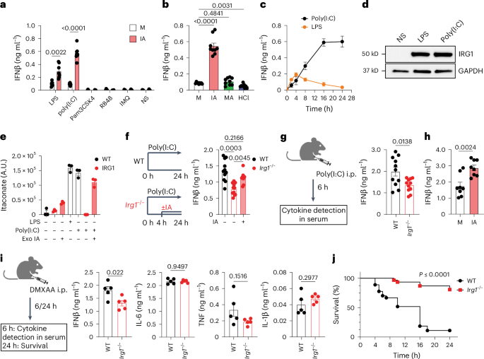
Looking through the candidate list again, I think there's an interesting thread that ties several of the top genes together, which is regulation of type I (alpha/beta) interferon signaling.

OLFM4
Vandenberghe-Dürr et al. said:
LL-37-DNA complexes induced strong IFN-α production by pDCs, which was significantly inhibited by pre-incubation of LL-37 with increasing concentrations of OLFM4
No directionality info from eQTLs.

PEBP1
Candidate Genes said:
Link to disease: Positive feedback loop between PEBP1/RKIP and TANK-binding kinase 1 that is essential for type I interferon production in anti-viral innate immunity (49). Induces HIV-1 latency and is induced by IFN signalling (50).
Candidate Genes said:
The allele that increases the risk of ME/CFS is associated with increased PEBP1 expression in the tibial artery.

TRIM38 (type I interferons are one of the main products of TLR and cGAS-STRING/viral RNA sensing)
Molecular function: Expression of TRIM38 is induced by viral infection, type I interferons and Toll-like receptor (TLR) ligands. Negatively regulates TLR3/4 signalling by targeting TRAF6 and NAP1 for proteasomal degradation (34). TRIM38 is a SUMO ligase for both cyclic GMP-AMP synthase and STING, molecules that regulate the innate immune response to DNA viral infection, which prevents their degradation (35).
Cellular function: Negative regulator of the innate immune and inflammatory responses triggered by viral RNA, LPS, and TNF-α and IL-1β. Prevents the excessive response to DNA viral infection and uncontrolled inflammation (35)
Candidate genes said:
The allele that increases the risk of ME/CFS is associated with increasing or decreasing TRIM38 gene expression, depending on tissue.

KLHL20
Candidate genes said:
Molecular function: Substrate-specific adapter of a BCR (BTB-CUL3-RBX1) E3 ubiquitin-protein ligase complex involved in interferon response and anterograde Golgi to endosome transport.
Candidate genes said:
The allele that increases the risk of ME/CFS is associated with decreasing KLHL20 gene expression
E3-ubiquitin ligases are typically associated with degradation of interferon, but it might not be so cut and dry.

ZNFX1
Candidate genes said:
An interferon-stimulated dsRNA sensor that specifically restricts the replication of RNA viruses early after infection (82). Studies are unclear whether ZNFX1 is localised to the mitochondrion.
I'll note that though this is an interferon-stimulated gene, it's activity as a dsRNA sensory would also trigger interferon.
Candidate genes said:
The allele that increases the risk of ME/CFS is associated with increased ZNFX1 expression in blood and brain tissues.

And interestingly, PRDX6, a cousin of PRDX5--which is inhibited by itaconate to allow type I interferon production in macrophages
Candidate genes said:
The allele that increases the risk of ME/CFS is associated with decreasing PRDX6 gene expression.

The BTNs could also be part of the story via their regulatory effect on TLRs. Plus TLR/type I interferon signaling utilizes MAPK signaling, which potentially implicates ZNF322 (increased) and SUDS3 (increased).Plus PTGIS (decreased expression), since prostaglandins are known to inhibit interferon signaling in some contexts.

Just throwing things at a wall to see what sticks based on the assumption that at least some of these genes are actually affected by the identified SNPs. The directionality of most of these (where eQTL data is provided) seems to generally be in the direction of enhanced interferon in ME/CFS (either increased expression in genes that are positive regulators or decreased in genes that are negative regulators), so that would be some weak evidence against the idea that these SNPs are driven by [edit: poorer protection in ME/CFS cases] against viral infections.

[Edit: and acknowledging that I’m probably biased here since I already think there’s a good case for type I interferons in ME/CFS]
 
Last edited:
Back
Top Bottom