Central noradrenergic deficiency in post-infectious chronic fatigue: neurobehavioral correlates, 2026, Aregawi et al

Age:
The Parkinson’s disease group was older than the other groups, with a mean age of 64.1 years (SD 10.6; ≈<0.0001 by Tukey’s test), and the healthy volunteer group was older than the PI-ME/CFS group (49.2 vs 38.7 years; P=0.027).


Screenshot 2026-05-24 at 7.30.34 PM.png

Those mean ages are different. From youngest to oldest
ME/CFS (38.7) > PASC (45.2) > HV (49.2) > PD (64.1)

The Mean NE Pathway Index order from lowest to highest with approximate mean indices
PD (45) > ME/CFS (53) > PASC (55) > HV (65)
The differences in the pathway index between the ME/CFS, PASC and HV groups aren't actually enormous.

We know the PD cohort has a problem with the NE Pathway. Putting the PD group aside, the correlation of increasing age with increasing index might explain the findings.

From what I can see from he literature, NE levels in the CSF increase with age, both in men and women.

Increased plasma and cerebrospinal fluid norepinephrine in older men: differential suppression by clonidine
To evaluate the effect of advanced age on central nervous system noradrenergic activity, cerebrospinal fluid (CSF) and plasma norepinephrine (NE) concentrations were measured concurrently in 14 older [mean, 65 +/- 9 (+/- SD) yr] and 33 younger (25 +/- 2 yr) normal men. CSF NE was significantly higher in older men than in young men [214 +/- 75 (+/- SD) vs. 164 +/- 56 pg/mL (1.26 +/- 0.44 vs. 0.97 +/- 0.33 nmol/L)
Look at that difference. 164 for the group with an average age 25, and 214 for the group with an average age of 65.


Cerebrospinal fluid norepinephrine and cognition in subjects across the adult age span
Adequate central nervous system noradrenergic activity enhances cognition, but excessive noradrenergic activity may have adverse effects on cognition. Previous studies have also demonstrated that noradrenergic activity is higher in older than younger adults.
That paper confirms that multiple papers have fund that NE levels and NE activity increase with age. They found that, after adjusting for age, higher levels were correlated with poorer cognitive performance. Just possibly the ME/CFS and PASC participants in the 2026 paper that is the subject of the thread actually have NE levels consistent with good cognitive levels, i.e. these were mostly smart people who were able to get themselves into the NIH studies.
Noradrenergic system activity appears higher in older compared with younger adults, both peripherally and in the central nervous system (Featherstone et al., 1987; Lawlor et al., 1995; Supiano et al., 1990). Our group has previously demonstrated that concentrations of cerebrospinal fluid (CSF) norepinephrine (NE), the primary noradrenergic neurotransmitter, are higher in older compared with younger adults (Elrod et al., 1997; Raskind et al., 1999).
CSF NE concentrations were higher in older participants (regression coefficient = 0.89 [average difference in CSF NE concentration (picogram per milliliter) for each year difference in age], standard error (SE) 0.27, p = 0.001; Fig. 2). Notably, there was a wider range in CSF NE values in older compared with younger adults. This age-related difference in CSF NE did not differ by gender (age by gender interaction, p = 0.60).

I also saw something about menopause causing increases in NE levels, something about the withdrawal of oestrogen.

So, I don't think that this study provides evidence of noradrenergic activity deficiency in ME/CFS and PASC. I haven't finished reading the paper yet, but I think what we are seeing here is probably mostly just what we would expect given the differences in age. The ME/CFS and PASC people might in fact have very healthy NE activity for their ages.

I wonder if the authors plotted individual ages against individual NE activity?
 
Last edited:
Sex:
The groups were similar in sex composition and body-mass indices (Table 1).
The authors claim that the groups were similar in terms of sex ratios. But, they absolutely were not. there is a P value there and it is significant.

Percentage female by group:
HV: 32.5%
PASC: 67.6%
PD: 37.5%
ME/CFS: 56.3%

The PASC percentage of females is more than twice as big as the HV percentage of females. It is not okay to say that the groups are similar in terms of sex composition - they are not.

Although the healthy volunteer and patient groups differed in sex makeup, analysis of covariance showed that sex did not influence the difference between the healthy volunteer and patient groups in values for the Norepinephrine Pathway index (data not shown).
Fair enough that they bothered to check if the sex ratios made a difference to the results, and in the case of the NE activity, probably they don't. But, they did no similar analysis of the impact of age on NE activity. I think that is because it would undermine their claim of noradrenergic activity deficiency.


I've now read the paper apart from the supplementary materials. I think this paper is a bit rubbish, mostly due to the poor control matching. I suspect at least some of the authors know it is. In the Limitations they say
This study involved relatively small cohorts studied in a highly comprehensive manner at a single center. Generalizability of the results to the broader populations of PI-ME/CFS and PASC patients is unknown.
CSF levels of catecholamines and their metabolites vary extensively across individuals.
However, they say nothing about the problem with the poor matching of age.

They go on about the noradrenergic activity deficiency in the ME/CFS and PAIS cohorts. I doubt that there is a deficiency. I think we would need to find a significant difference between the actual NE and metabolite values and those for healthy people of the same age in order to substantiate the suggestion in this paper.
 
Another is the need for our usual task of checking the demographics of the cohorts in studies like these. i.e. male/female, ages, pre-menopausal/post-menopausal, time of the menstruation cycle, time of day of sample collection, sedentary versus more active. The latter could possibly explain the correlation between hand grip and button clicking performances with norepinephrine levels.
Yes, I was surprised that activity levels and/or deconditioning were not mentioned in the Aregawi paper. But I have found it hard to get a clear picture of how low activity levels and, conversely, exercise, affect norepinephrine levels.

From what I can find, people who are sedentary have lower norepinephrine levels than people who exercise, and their system (/anyone's with low norepinephrine) might overreact to challenges.

Acute exercise results in temporarily higher norepinephrine, but regular exercise lowers norepinephrine. (Or does it bring it closer to the "right" level? Unclear.)

Clonidine lowers norepinephrine levels, and in a trial it made pwME worse.

So if we think that both regular exercise and clonidine often make pwME feel worse, we could speculate that lowering already-low norepinephrine is not a good idea, and the low norepinephrine of pwME is not due to their low activity levels.

Maybe.

Who would know more about the effect of exercise on norepinephrine?
 
poor matching of age
I agree.

In the ME/CFS intramural study, there was closer matching of age (42 ± 13 versus 38 ± 15 years), so I guess we'd have to explain why norepinephrine correlated with grip strength and EEfRT there.

I don't think I had clocked/internalised this before, though, in the Supplementary Info:
Characteristics of Individuals who Underwent a Second Study Visit
All participants were invited to return for a second study visit to measure exercise response.
Eight PI-ME/CFS and nine HVs were able to return for participation prior to the study being
closed due to the SARS-CoV-2 pandemic. The PI-ME/CFS participants (age: 42±13 years, female:
50%, BMI: 26.7±5.0) were older and more often male than those that could not participate
(age: 34.5±13.8 years, female %: 78%, BMI: 24.8±5.4). [bolding added]

Those are the participants who did the CPET, and they had higher baseline CSF norepinephrine than the bigger group. That could support your age argument @Hutan as being the differentiating factor, rather than "effort preference". But the sex difference is huge too. The suggestion that "strong encouragement" in the CPET is why "effort preference" did not correlate with CPET is absurd when there are such big differences between the whole group and the subgroup that did CPET.

Studies with tiny samples need particularly careful matching.
 
In the ME/CFS intramural study, there was closer matching of age (42 ± 13 versus 38 ± 15 years), so I guess we'd have to explain why norepinephrine correlated with grip strength and EEfRT there.
Is that age data specifically reported for the norepinephrine study?

The intramural study paper (Deep Phenotyping) said
In cerebrospinal fluid, the PI-ME/CFS group had statistically significant decreased levels of DOPA, DOPAC, and DHPG (Fig. 6a–c). Levels of norepinephrine, cys-DOPA, and dopamine did not differ between the groups. These results did not change after excluding data from parti-cipants taking central-acting medications. Additional analysis revealed that norepinephrine correlated with both time to failure and effort preference in PI-ME/CFS participants (Fig. 6d, e).
So, notably, they didn't find lower levels of norepinephrine in that study.

Presumably the n=16 PI-ME/CFS are from the Intramural cohort under second author Brian Walitt.

If the data for norepinephrine is the same for the ME/CFS group, then it must only be the addition of the NE metabolites that allow the authors to make the claim of noradrenergic activity deficiency in this paper? Perhaps we need to look at the data for the metabolites a bit more.

(I'm not sure it is worth trying to unravel the EEfRT result? That study was such a debacle in so many ways.)
In my next post I'll look at bit closer at the NE vs time to failure result in the ME/CFS intramural study.
 
Last edited:
If the data for norepinephrine is the same for the ME/CFS group, then it must only be the addition of the NE metabolites that allow the authors to make the claim of noradrenergic activity deficiency in this paper?
Yes this was my assumption based on the way they presented the results. They never reported NE by itself (as far as I remember anyway), and instead included an argument about why NE + metabolites made more sense as a measure. No idea of the validity of that. One of the authors does seem to be practically an emeritus prof of NE so maybe that lends some credibility?

I'd love it if low NE turnover was our problem, or part of it, because it seems relatively tractable. But OTOH the point you made about age does seem like it could wipe out the whole difference.
 
Looking at the claimed CSF NE levels versus the time to failure in the ME/CFS intramural study:

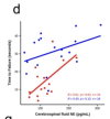
They are suggesting that levels of norepinephrine in the ME/CFS group correlated with time to failure .
Figure 6d
Screenshot 2026-05-26 at 3.24.21 PM.png


Figure 6d is the chart of NE levels and time to failure. At first glance, that result seems odd - we would expect higher NE levels to be associated with older people, and older people to tend to have lower times to failure. But, potentially there are so many confounders there - for a start we don't know which datapoints belong to male or female participants - I don't think there was any adjustment for sex. The reliability of the relationships are low, and probably heavily influenced by a couple of outliers. And, there is the potential impact of the ME/CFS people being in various levels of PEM at the time of the strength assessment.

I'm not sure that it is worth spending more time trying to understand that result - unless we can find out more about the age and sex associated with each individual data point.

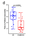
Figure 3d
Screenshot 2026-05-26 at 3.22.31 PM.png
(d) time to failure of HV (blue; n = 18 independent participants) and PI-ME/CFS (red; n = 16 independent participants), unadjusted two-sided t-test for independent samples with equal variance, p = 0.0002.

I thought it was worth checking the data from the figure 3d from the ME/CFS intramural study paper.

Looking at the healthy controls, I note that 20 controls provided grip strength data, but only 18 provided time to failure data. So, the data for two controls is missing.

Also, have a look at the time to failure for the bottom three control data points in Figure 3d. One is just less than 10, one is less than 15 (maybe 13?) and one is maybe 17.5. But, look at the data points in Figure 6d. The bottom data point looks to be where it should be. But, the second lowest looks more like 17 than 13. And the third lowest looks more like 19 than 17.5. I don't think there has been adjustment for sex or something, because all the other data points look pretty accurate. So, what happened with those two data points?

Could someone check what I've said there is right - that the second and third lowest control points appear to change between figures 3d and 6d?

Looking at the ME/CFS group, I note that 16 ME/CFS provided time to failure data, but only 15 are included in the 6d chart against NE. I guess it's possible that the 16th ME/CFS person couldn't provide a CSF sample. However, the missing value is the second highest time to failure, maybe 29 seconds. If that value had been plotted on the chart, maybe it could have made the relationship not significant.

That's the problem with these tiny cohorts - the results are so vulnerable to a couple of data points being dropped out, and to confounders such as sex and age.
 
Last edited:
I find it hard to square low noradrenaline with the claimed link to POTS, which, together with ME/CFS in general has in the past been claimed to indicate upping of sympathetic and downing of parasympathetic I think. But that was likely nonsense anyway.
@Jonathan Edwards have you read my analysis on orthostatic intolerance in UARS? (Sleep-disordered breathing (UARS/OSA) and orthostatic intolerance). Parasympathetic hyperactivation in response to effortful inspiratory flow limitation is a proposed driver:
It found that in UARS patients, abnormal parasympathetic responses occurred specifically during flow-limited breaths with increased respiratory effort rather than during flow limitation alone -- pointing to negative intrathoracic pressure as a key variable. The authors propose that nightly parasympathetic/vagal hyperactivation during effortful IFL may, over time, produce functional sympathetic de-innervation -- a blunting of the sympathetic nervous system's ability to constrict blood vessels -- that has been demonstrated experimentally using specific breathing manipulations during wakefulness. If chronic vagal hyperactivation progressively impairs sympathetic vasoconstrictor responses, the ability to maintain blood pressure on standing would be compromised.

ANP-mediated hypovolemia and baroreflex downward resetting have been proposed as additional candidate mechanisms on the basis of the known physiological effects of sustained negative intrathoracic pressure, and are consistent with the resting tachycardia and orthostatic hypotension pattern observed by Guilleminault et al. (2001).
Based on my personal experience, I find it plausible that hyperadrenergic POTS in some patients is a compensatory response to whatever is actually going on with the orthostatic intolerance across the board (hypovolemia, functional sympathetic de-innervation, etc.)
Of note, my OI started out with more purely OH-type symptoms (lightheadedness/losing vision on standing) early on and I developed more (hyper) POTS-type symptoms over time. I’ve had both OH and POTS on NASA lean tests (even within short time frames - e.g. one time I had hyperPOTS one day when I had overexerted and had the “running on adrenaline” feeling and OH the next day in PEM).

Low norepinephrine is actually consistent with the sleep-disordered breathing paradigm. This study (Implications of sympathetic activation for objective versus self-reported daytime sleepiness in obstructive sleep apnea, 2022, Chen et al) found that OSA patients with purely subjective sleepiness (i.e. fatigue -- high Epworth Sleepiness Scale scores, but don't actually fall asleep quickly; I've shared with you before that the Epworth Sleepiness Scale measures a mix of fatigue and objective sleepiness, which are uncorrelated symptoms in OSA patients) had lower 24-hour urinary norepinephrine than OSA patients with no subjective or objective sleepiness (OSA patients with objective sleepiness had the highest 24-hour urinary norepinephrine). EDS = excessive daytime sleepiness.
Notably, subjects with purely self-reported EDS also had significantly lower 24-hour urine norepinephrine than those with no self-reported or objective EDS, supporting that there are different physiological processes underlying objective and subjective EDS in OSA.
Moreover, patients with OSA with purely self-reported EDS (34.03 ± 7.09 μg) also had significantly lower 24-hour urinary norepinephrine compared with those with both self-reported and objective EDS (53.09 ± 4.53 μg, p = 0.024) and those with no EDS (54.95 ± 4.62 μg; p = 0.016). These results remained similar when adjusted for minimum oxygen saturation instead of AHI. In sensitivity analyses after excluding patients taking anti-hypertensive medications, the findings remained similar.
From a write-up I'm working on (don't worry, I'll elaborate on the Phenotype 1 vs. 2 framework more in that, but Phenotype 2 is the fatigue subtype proposed to be driven by a physiological stress response to inspiratory flow limitation as theorized by Dr. Gold -- theory summary here):

Within this framework, the finding of Chen et al. (2022) that OSA patients with purely subjective excessive daytime sleepiness (EDS) had significantly lower 24-hour urinary norepinephrine than both objective EDS patients and patients with no EDS at all may be consistent with the same effortful inspiratory flow limitation-driven autonomic imbalance proposed to underlie low BP/orthostatic hypotension (OH) in UARS operating subclinically across a broader Phenotype 2 (UARS/OSA) population. That these patients show low 24-hour urinary norepinephrine despite OSA diagnosis is not necessarily inconsistent with the BP divergence observed between UARS and OSA patients in Guilleminault et al (2001): hypoxemia-driven pressor mechanisms in OSA may be sufficient to prevent overt low BP/OH even in the presence of reduced sympathetic tone. The autonomic signature of Phenotype 2 may represent a spectrum from subclinical sympathetic attenuation detectable only biochemically, through orthostatic intolerance, to frank orthostatic hypotension.
In UARS, those pressor responses are largely absent, leaving these mechanisms unopposed. The AHI 5 cutoff may be coincidentally approximating the threshold at which intermittent hypoxia-related effects becomes sufficient to counteract hypotensive mechanisms at a population level — explaining why low BP and OH specifically, rather than OI in general, appear most distinctively associated with UARS rather than OSA.
 
Last edited:
Yes this was my assumption based on the way they presented the results. They never reported NE by itself (as far as I remember anyway), and instead included an argument about why NE + metabolites made more sense as a measure.
Yes, I've criticised other papers for presenting us with an index but not presenting the values of the things that get put into the index. It's annoying, especially in cases like this where there aren't a lot of features, and the index is made especially for the paper:

To analyze the neurochemical data we devised, validated, and applied novel indices of central dopaminergic and noradrenergic pathway activities.
We wished to summarize the neurochemical information by validating and applying overall indices of central dopaminergic and noradrenergic function that readers could comprehend.

Not giving us the levels of the inputs to the index means that we are not able to check whether the reported levels and ratios are within normal bounds even though they might differ from a small control cohort. I don't think providing that data would have made the paper difficult to comprehend.


I think they possibly intended to give us the data for each catecholine and metabolite - the text suggests the data should be there:
Individual values and group mean levels of CSF catecholamines and metabolites by group are shown in Fig. 2.
Instead, the charts are just for the indexes.


CSF MHPG levels were lower in the PI-ME/CFS (12.6 [4.6, 20.6], P=0.00044), PASC (7.8[1.4,14.3], P= 0.01035), and Parkinson’s disease (16.0 [9.7, 22.3], P<0.0001) groups compared to the healthy volunteer group.
They don't tell us about the NE or the DHPG levels. Nor do they tell us what the MHPG levels and variance were in the healthy volunteers. They don't even tell us the units.

I've checked the supplementary material. It isn't good enough to be presenting this story of deficiency in a group of people when the data and the presentation is so poor, and the index has not been properly validated on a large sample of healthy controls.
 
Last edited:
The literature about MHPG is not robust. If you think you have some fact, read another study, because it will probably say the opposite. One reference about brain chemicals including MHPG says:
The literature in this field is not only scarce, it also almost exclusively reports data from patients with (suspected) neurological disease [25, 26, 28, 34] due to the burden of a lumbar puncture necessary to obtain CSF. Furthermore, most studies were conducted more than twenty years ago and only involved analysis of one or a few amines

I wonder about the 'healthy volunteers' in this Aregawi study. All we are told is that these were people who had come into the NIH for other studies. It is not clear if they were also healthy controls for those studies.

A study on MHPG - found that MHPG levels increased with age:
We have investigated neurotransmitter-related markers of the cerebrospinal fluid (CSF) in a carefully screened series of normally aging subjects in standardized conditions in order to find out the influence of age and other confounding factors on CSF measures. The levels of 3-methoxy-4-hydroxyglycol (MHPG) and the activity of acetylcholinesterase (AChE) also increased with age, while homovanillic acid (HVA), 5-hydroxyindoleacetic acid (5 HIAA) and immunoreactivities of somatostatin (SLI), beta-endorphin (BLI) and adrenocorticotropic hormone (ACTH) were unrelated to age. The gender of subjects had no significant effect on the levels of neurotransmitter markers, while seasonal changes, as well as height and weight of the subjects seemed to cause some variations in the levels of HVA, dopamine-Β-hydroxylase (DBH) and ACTH. The study underscores the importance of standardized conditions and matched patient groups in the CSF studies.

One study (the Tiigchelaar 2023 study) suggests that MHPG, unlike NE, may be able to cross the blood brain barrier, as they found a correlation between plasma and CSF levels. I'm not sure what that means for the Aregawi study's idea of making an index to reflect brain noradrenergic activity

Another study confirms that:
Interestingly, MHPG easily passes the blood–brain [32] and blood-CSF [33] barrier.
A more recent study confirmed that MHPG, unlike NA [20], passes the blood–brain, as well as the blood-CSF barrier [61],

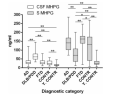
Here are some values for MHPG from that study. The controls might possibly be a bit dubious when it comes to the 'healthy' part. Healthy people tend not to want to undergo a lumbar puncture.

Screenshot 2026-05-26 at 6.54.05 PM.png
The white boxes on the left are for CSF, the grey boxes on the right are for serum. The first three categories are for types of dementia (including Parkinsons Disease dementia) and then there are controls and young controls. Clearly, there is a lot of variation, even in healthy controls. The age range for the young controls was 17 to 50, and so probably is indicative of what could be expected for healthy people in the age range of the Aregawi PASC and ME/CFS cohorts. The age range for the controls was 50 to 93 (what they were doing giving a 93 year old a lumbar puncture, I do not know).

Values tend to increase with age (but there is a lot of variation). So, as for NE, it's not surprising that the ME/CFS and PASC cohorts had lower levels than the controls.

I'll stop now. This is such a frustrating paper. Perhaps there is something interesting about the noradrenergic systems of people with ME/CFS. But, the deeply inadequate presentation of data in this paper is not providing any convincing evidence of it.
 

Attachments

  • Screenshot 2026-05-26 at 6.11.52 PM.png
    Screenshot 2026-05-26 at 6.11.52 PM.png
    511.9 KB · Views: 0
At first glance, that result seems odd - we would expect higher NE levels to be associated with older people, and older people to tend to have lower times to failure.
Not sure we'd necessarily expect that. We know that a 40 year difference was significant in the paper you quoted above. We don't know that patients and controls differing by about 4 years would be significantly different based on age alone.

They never reported NE by itself (as far as I remember anyway), and instead included an argument about why NE + metabolites made more sense as a measure. No idea of the validity of that.
Not giving us the levels of the inputs to the index means that we are not able to check whether the reported levels and ratios are within normal bounds even though they might differ from a small control cohort. I don't think providing that data would have made the paper difficult to comprehend.
I agree, this is problematic. They say:
the central noradrenergic index we used was dominated by CSF MHPG

I have been reading that MHPG differs quite a lot by sex, with females having lower levels. That makes the non-matching for sex potentially problematic, because the ME/CFS and PASC groups had higher percentages of females than the PD & HV groups.

MHPG is low in a lot of conditions, including depression. Found a few papers by a researcher called Garvey who looked at this a lot in the 90s e.g. Do Low Levels of MHPG in Depressive Spectrum Patients Normalize after Successful Treatment? Treatment was with "imipramine, cognitive therapy, or a combination of the two". Imipramine is a tricyclic that increases norepinephrine. There was no placebo pill. This struck me:
DSD patients had no significant change in the low levels of baseline MHPG following symptom improvement. In fact, the low levels of pretreatment MHPG were even lower after treatment, though not significantly so. This data is consistent with a hypothesis that the low MHPG found at baseline in DSD patients may be a trait phenomenon. If the low level of MHPG in DSD patients is a biological trait, this trait could be part of the pathogenetic mechanism or vulnerability for DSD depression.

I don't know if that was replicated or is true of other drugs that increase NE. But if so, and if that were also true in ME/CFS, then you could have a situation where researchers try to manipulate NE levels and come up short, because you can't fix a predisposition.

My concern is that I think we may be headed for a trial of a norepinephrine modulator in ME/CFS (akin to Cymbalta/Savella for fibromyalgia).

Why do I think this? This is from Walitt et al. 2024 aka the ME/CFS intramural study:
This model suggests places for potential therapeutic intervention and explains why other therapies have failed. The finding of possible immune exhaustion suggests that immune checkpoint inhibitors may be therapeutic by promoting clearance of foreign antigen. Immune dysfunction leads to neurochemical alterations that impact neuronal circuits, which may be another point of intervention. Therapeutically targeting downstream mechanisms, with exercise, cognitive behavioral therapy, or autonomic directed therapies, may have limited impact on symptom burden, as it would not address the root cause of PI-ME/CFS. However, combination therapy affecting multiple pathways could be considered. The finding of substantial physiological differences related to sex suggest that there may not be a single unified mechanism that leads to PI-ME/CFS and that successful therapy may require a personalized medicine approach.


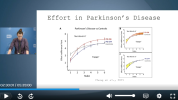
In the symposium, Dr Madian presented this slide showing figures from Chong et al. 2015, saying how similar these findings were to those in ME/CFS. What Chong et al. 2015 found was that people with Parkinson's had reduced reward motivation, which was reversed (and then some) with dopamine-raising medication.

1779780423785.png


The implication seemed to be that there may be a medication that can reverse pwME's supposed "effort preference" (which did not entail reduced reward motivation, and was not calibrated to ability). Even if we change that to "a medication that can reverse pwME's lower norepinephrine levels", we've got at least two problems:

1. Are they really lower? These are small samples - in the case of ME/CFS, tiny - and the groups differ in baseline characteristics. See @Hutan's point above that the group differences in NE levels in the Aregawi study could be caused by group differences in age.

2. If Aregawi et al.'s findings were replicated in larger, better-matched samples, then we'd still have the issue of significant overlap with healthy volunteers.


To check whether the "central noradrenergic deficiency" is "pathophysiologically significant", Aregawi et al. 2026 suggest "examining effects of manipulations of central norepinephrine release, reuptake, or metabolism on neurobehavioral measures". That would have to be placebo-controlled. But importantly, if it did improve performance, we would need to know that people can tolerate the consequences of increased activity long-term, rather than deteriorating 3/6/12/24 months down the line.

I have not seen anything yet that would convince me that a norepinephrine-modulator will be a gamechanger in ME/CFS like levodopa is in Parkinson's. And levodopa's effect on reward motivation in Parkinson's has downsides: gambling etc.
 
Last edited:
Is that age data specifically reported for the norepinephrine study?
Looking into that further. That's all recruited participants in the intramural study. N would be 39 based on the fact that they break down sex as 10 versus 6 men; 12 versus 11 women, but n=38 based on main paper saying 17 pwME and 21 HVs. N=34 provided CSF NE data (16 pwME and 18 HVs). So one patient missing and 3 controls missing.
 
Just noticed that in the intramural study, 5 healthy volunteers provided CSF NE but did not do the EEfRT task, and their mean baseline CSF NE was 118.6 compared to 156.8 for the HVs who did do the EEfRT. (Mistakes possible, not double-checked.) 2 patients provided CSF NE but did not do the EEfRT. One's NE was 90 (patterning with the poor completers); the other was 139 (patterning with the good completers).

If CSF norepinephrine had a real influence on performance in the EEfRT, then that's problematic. Would there still have been group differences in the EEfRT if all healthy volunteers had done it?
 
Central noradrenergic deficiency in post-infectious chronic fatigue: neurobehavioral correlates

Aregawi, Lillian; Walitt, Brian; Sullivan, Patti; Norato, Gina; Benjamin, Rohit Ninan; Goldstein, David S

Abstract
Fatigue, brain fog, and post-exertional malaise are major features of post-infectious myalgic encephalomyelitis/chronic fatigue syndrome and post-acute sequelae of severe acute respiratory syndrome coronavirus 2. To date, no specific neurotransmitter abnormalities have been found in either condition.

We examined the possibility of central catecholaminergic involvement and clinical correlates in post-infectious myalgic encephalomyelitis/chronic fatigue syndrome and post-acute sequelae of severe acute respiratory syndrome coronavirus 2 groups compared to healthy volunteers and, as positive controls, Parkinson’s disease patients. In an observational, cross-sectional cohort study conducted at the National Institutes of Health Clinical Center we measured CSF levels of catecholamines and metabolites in the four groups and assessed correlations with neurobehavioral measures.

CSF indices of the central Norepinephrine Pathway (norepinephrine+3,4-dihydroxyphenylglycol+3-methoxy-4-hydroxyphenylglycol) and Dopamine Pathway (dopamine+3,4-dihydroxyphenylacetic acid+homovanillic acid) were measured and related to patient-recorded outcomes and physiological assessments.

Mean values for Norepinephrine Pathway activity (in pmol/mL) were lower in the post-infectious myalgic encephalomyelitis/chronic fatigue syndrome, post-acute sequelae of severe acute respiratory syndrome coronavirus 2, and Parkinson’s disease groups compared to the healthy volunteer cohort (post-infectious myalgic encephalomyelitis/chronic fatigue syndrome (-15.9, 95% confidence interval [-26.1, -5.7], P=0.0006); post-acute sequelae of severe acute respiratory syndrome coronavirus 2 (-9.62, [-17.9, -1.4], P=0.015); Parkinson’s disease (-19.4, [-27.5, -11.3], P<0.0001)). Post-acute sequelae of severe acute respiratory syndrome coronavirus 2 participants with post-exertional malaise had evidence of central noradrenergic deficiency compared to healthy volunteers (-18.3 [-31.3, -5.3], P=0.0018).

The post-infectious myalgic encephalomyelitis/chronic fatigue syndrome and post-acute sequelae of severe acute respiratory syndrome coronavirus 2 groups did not differ from the healthy group in values for the Dopamine Pathway index. Across all participants, Norepinephrine Pathway activity correlated positively with handgrip duration and general health and negatively with fatigue.

We conclude that post-infectious myalgic encephalomyelitis/chronic fatigue syndrome and post-acute sequelae of severe acute respiratory syndrome coronavirus 2 feature a specific central neurotransmitter pattern involving noradrenergic but not dopaminergic deficiency. The noradrenergic abnormality is associated with major symptoms such as post-exertional malaise.

Web | DOI | PDF | Brain Communications | Open Access
I think the study topic (role of norephinephrine in ME/CFS, PASC etc) is exciting - given what NE does in the brain.

If the results hold up (which I cannot assess), they would not be specific to ME/CFS as the comparison with PD shows.

What worries me: Here are world experts in dysautonomia, neurophysiology and whatever. And then their only explanation for the apparent NE deficit is „ATP deficiency“. What about messed up locus coeruleus regulation (e.g., LC function is intricately interwoven with the functions of the Anterior Cingulate Cortex, frequently implicated as a player in ME/CFS)? What about lost synchrony with „demand“ signals (like: stressful events)? What about this being reflective of dysregulation in other systems working together with or influencing the locus coeruleus (the vasopressin and the orexin systems come to mind – apropos: would be nice if they had some CSF left to assess orexin levels – apparently never done in ME/CFS).

If they´d be really excited about their own results they´d probably have provided a more exciting discussion.
 
What I find very interesting is that nicotine patches helped me by far the most personally and those do increase norepinephrine. Aripiprazole can also modulate its release and has anecdotally helped many patients. I've got some friends on almost full recoveries from LDA.


Highly anecdotal ofc but makes me want to see proper trials on both substances.

At least in the one retrospective study on LDA from Stanford results looked cautiously promising:

 
Last edited:
What I find very interesting is that nicotine patches helped me by far the most personally and those do increase norepinephrine. Aripiprazole can also modulate its release and has anecdotally helped many patients. I've got some friends on almost full recoveries from LDA.


Highly anecdotal ofc but makes me want to see proper trials on both substances.
Drugs like venlafaxine (Effexor) and duloxetine (Cymbalta) also increase norepinephrine, but I don't know that people with ME/CFS find them helpful for their ME/CFS (as opposed to for pain, depression or anxiety).

Some ADHD meds increase norepinephrine too. Again, it could be hard to disentangle positive impact on ADHD from impact on ME/CFS.

The Aregawi paper mentions atomexetine and cites this Alzheimer's trial. Maybe they consider it a possibility for us?
Yea I haven’t heard great things about drugs from that category. Maybe it’s about the different ways in which these drugs affect the levels. Neither Nicotine nor LDA do it directly or by inhibiting reuptake so maybe there’s the catch.
 
CSF MHPG levels were lower in the PI-ME/CFS (12.6 [4.6, 20.6], P = 0.00044), PASC (7.8 [1.4, 14.3], P = 0.01035), and Parkinson’s disease (16.0 [9.7, 22.3], P < 0.0001) groups compared to the healthy volunteer group.
I haven't read this thread's paper or the following paper in detail yet, but just noting that it's another study that found low MHPG in the CSF:

Plasma and cerebrospinal fluid monoamine metabolism in patients with chronic fatigue syndrome: Preliminary findings (Demitrack et al., Biological Psychiatry, 1992)
[...] 19 patients meeting CDC research case criteria for chronic fatigue syndrome and in 17 normal individuals. Patients with chronic fatigue syndrome showed a significant reduction in basal plasma levels of MHPG and a significant increase in basal plasma levels of 5-HIAA.
 
W/r/t "explanation for apparent NE deficit is ATP deficiency

This is a backdoor look at the issue, but...


Thiamine deficiency depletes cortical norepinephrine and impairs learning processes in the rat​


Is the thiamine deficiency depleting cortical norepinephrine due to reduced ATP?

Edit in for possible relativity:

High-dose thiamine as initial treatment for Parkinson's disease​


And also,

ATP-sensitive and Ca2+-activated K+ channel activities in the rat locus coeruleus neurons during metabolic inhibition​

The ATP level also affects the locus coeruleus?

 
Last edited:
Back
Top Bottom